Every year, hundreds of thousands of infants around the world arrive far too early, and for them the womb is traded, sometimes months before their due date, for the humming, brightly lit, instrument-lined environment of the neonatal intensive care unit. Modern neonatal medicine has transformed survival at extremely low gestational ages, and that success has created a question that medicine is only now beginning to confront in earnest: what is it actually like, chemically and developmentally, to grow inside a hospital? A new commentary by Julia A. Bauer and Joann Romano-Keeler, published in the Journal of Exposure Science & Environmental Epidemiology, argues that the NICU should be understood not merely as a lifesaving clinical space but as a distinct developmental exposome, a complete constellation of nutritional, chemical, physical, and sensory exposures that replaces the intrauterine environment during a period of extraordinarily rapid brain growth, organogenesis, and physiological maturation.
The concept of the exposome, the cumulative measure of environmental exposures an organism experiences across its lifespan, has gained traction in adult epidemiology, but its application to the newborn period carries a particular urgency. Very preterm birth fundamentally reshapes the developmental exposome by relocating a substantial portion of gestation outside the body. The fetus that would have been bathed in amniotic fluid, nourished continuously through the placenta, and shielded from most external chemicals instead receives parenteral nutrition through central lines, undergoes repeated blood transfusions, rests on plasticized medical devices, and breathes filtered, conditioned air. Each of these interventions is essential, and each introduces its own chemical signature. The commentary emphasizes that because survival has improved so dramatically, understanding how this prolonged hospital-based developmental setting influences long-term neurodevelopmental, respiratory, and cardiometabolic health has become a central question for children’s environmental health.
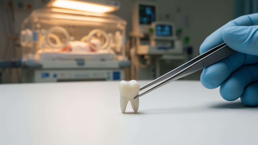
The central methodological obstacle has always been measurement. Collecting repeated biospecimens from medically fragile infants weighing sometimes less than a kilogram presents formidable practical and ethical challenges, and blood draws are limited by volume and clinical necessity. Bauer and Romano-Keeler highlight an elegant alternative that has been quietly maturing over the past decade: naturally shed deciduous teeth. Children lose their baby teeth in mid-childhood, and those teeth, it turns out, are meticulous biological recorders. As dentin mineralizes, elements circulating in the bloodstream are incorporated into the growing crystalline matrix in temporal sequence, layer by layer, much like tree rings. A distinctive microstructural feature called the neonatal line marks the moment of birth, giving researchers an exact chronological anchor from which to reconstruct week-by-week exposure histories stretching from fetal life through the entire NICU stay.
This approach offers something maternal biomarkers cannot. Measurements in maternal blood or hair primarily reflect maternal dose and may fail to capture placental transfer or fetal uptake, whereas prenatal tooth measurements derive from tissues formed in utero and therefore provide a more direct indicator of the fetal internal chemical environment. Postnatal metal exposure measured in teeth is increasingly investigated in cohort studies, but the exposures unique to the NICU have remained poorly characterized. The commentary accompanies a study in the same issue by Lieberman-Cribbin and colleagues that applies the technique to precisely this question, reconstructing weekly metal exposure histories using shed teeth collected from children across the United States who were born preterm.
The study drew participants from the Developmental Impact of NICU Exposures, or DINE, study, which enrolled children from multiple preterm cohorts participating in the Environmental influences on Child Health Outcomes, or ECHO, program. These infants spent a median of 85 days in the NICU, an extended hospitalization that allowed the researchers to examine metal exposure across a substantial portion of early development that would otherwise have occurred in utero. The findings were striking in their asymmetry. Several metals were higher on average during the prenatal period, while estimated average levels of lithium, rubidium, and strontium were higher postnatally, with the clearest difference observed for strontium. In other words, the chemical profile of development measurably shifts when the environment shifts from placenta to intensive care, and those shifts are written permanently into the mineralized tissue.
What could explain a postnatal rise in elements like strontium? The commentary lays out a set of plausible exposure pathways that the findings generate as hypotheses for future investigation. Infant feeding is an obvious candidate, since both human milk and formula carry distinct elemental profiles. Parenteral nutrition, the intravenous feeding that sustains the smallest infants for weeks, has a documented history of delivering trace metals, with earlier research identifying increased manganese deposition in the brains of infants receiving it and, in a landmark 1997 study, aluminum neurotoxicity in preterm infants fed intravenous solutions. Blood transfusions represent another under-recognized route, as red cell transfusions can carry toxic metals into a newborn circulation. Medical devices, including plasticized tubing and equipment, and other ambient features of the NICU environment round out the list. These pathways are especially relevant for extremely and very low birthweight infants, who receive the most prolonged and intensive nutritional and medical support precisely during a period of heightened developmental susceptibility.
Interpreting tooth biomarker data, however, demands careful attention to developmental biology, and the commentary is refreshingly candid about this complication. Temporal changes in elemental concentrations in dentin may reflect not only external exposures but also tissue growth, mineralization dynamics, shifts in nutrient requirements, and the maturation of metabolic and homeostatic pathways. A developing infant’s physiology is itself changing week by week, and those internal changes can alter which elements circulate and how they are deposited in mineralizing tissue. Distinguishing exposures arising from the external environment from endogenous developmental processes is therefore central to the enterprise. At the same time, the authors argue, the real value of these measurements lies in their ability to place both processes on a common developmental timescale, clarifying how the external environment and internal physiology jointly shape the chemical milieu of the developing child rather than pretending one can be isolated from the other.
The roadmap that emerges from the commentary is both practical and ambitious. Future studies that integrate tooth biomarkers with detailed clinical, nutritional, and environmental records will be essential for identifying the origins of NICU exposures: information on enteral and parenteral nutrition, medications, transfusions, respiratory support, medical devices, and treatment duration could help separate the chemical contributions of neonatal care from those of endogenous development. The authors also propose a structural innovation with lasting value: incorporating permission for future recontact and deciduous teeth collection into existing neonatal biorepositories, creating a resource that would enable longitudinal investigations of how early-life exposures influence health and disease for decades to come. Extending tooth-based methods beyond metals to plastic-associated chemicals and persistent organic pollutants, with potential applications to pharmaceuticals and other metabolites, would round out the picture of the NICU environment, building on emerging work that has already detected organic pollutants in primary teeth and linked fetal exposures to later outcomes.
The ultimate prize is causal clarity about long-term consequences. Preterm birth is associated with elevated risks of neurodevelopmental impairment, respiratory disease, and cardiometabolic dysfunction in adulthood, but the contribution of hospital-based exposures during the NICU course to those outcomes remains largely unmapped. Longitudinal follow-up of children whose teeth have been analyzed will be essential for determining whether exposures occurring during specific windows of the NICU stay are associated with later cognition, growth, respiratory health, or cardiometabolic function. If specific periods or specific care practices turn out to carry disproportionate chemical burden, the exposome map becomes a clinical instrument, pointing toward modifiable aspects of neonatal care, from nutritional formulations to device materials, that could be adjusted without compromising the lifesaving function of the unit itself.
What makes this research paradigm resonate beyond neonatology is its simplicity and its reversibility. Baby teeth that most families discard, or tuck into keepsake boxes, contain a week-by-week archive of one of the most chemically complex periods of human development, retrievable years later without a single invasive procedure. The NICU sustains life outside the intrauterine environment, and it does so brilliantly, but it is also a distinct setting in which clinical care, nutrition, environmental chemicals, and developmental physiology converge during a period of heightened vulnerability. Defining the developmental exposome of that setting, as Bauer and Romano-Keeler argue, may ultimately help design developmental environments that better support health across the entire life course, turning an underutilized biological resource into a bridge between the intensive care that saves infants and the lifelong health those survivors deserve.
Subject of Research: Reconstructing prenatal and NICU metal exposures in preterm infants using deciduous tooth biomarkers
Article Title: Defining the developmental exposome of the neonatal intensive care unit in preterm infants
Article References: Bauer, J. A., & Romano-Keeler, J. (2026). Defining the developmental exposome of the neonatal intensive care unit in preterm infants. Journal of Exposure Science & Environmental Epidemiology. https://doi.org/10.1038/s41370-026-00981-5
Image Credits: AI Generated
DOI: 10.1038/s41370-026-00981-5
Keywords: exposome, neonatal intensive care unit, preterm infants, deciduous teeth, metal exposure, biomonitoring, ECHO program, DINE study, parenteral nutrition, child environmental health, dentin biomarkers, strontium
News Source: Harold Sullivan. (October 8, 2026). Baby Teeth Reveal the Hidden Chemical World of the Neonatal Intensive Care Unit. Scienmag.