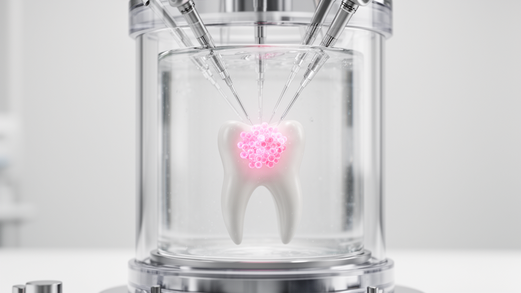
Published in BioData Mining, the study addresses a significant bottleneck in single-cell genomics: the manual and variable process of identifying specific cell populations within complex tissue datasets. By establishing a reproducible method for extracting uncultured MSCs, the team provides a tool that enhances the efficiency and scalability of analyzing dental pulp biology, particularly in the context of tissue repair and regeneration.
The primary challenge in analyzing human dental pulp single-cell RNA-sequencing data has been the reliance on manual verification of marker genes after unsupervised clustering. This traditional approach is time-consuming and subject to variability, as the choice of clustering resolution is often determined empirically. Such empirical decisions can lead to the under-segmentation or over-segmentation of MSC populations, or their mixing with other cell types. The study aimed to establish a computational workflow that automatically extracts these cells and investigates the characteristics of freshly-isolated MSCs using multiple public datasets.
The research team, led by Daigo Okada, Tomoko Takeda-Kawaguchi, and Ken-Ichi Tezuka, developed a workflow that identifies MSC populations based on a predefined marker set from a single-cell RNA-sequencing count matrix. The system systematically evaluates the performance of the marker set by optimizing the clustering resolution. The method calculates module scores for the specified MSC markers and selects the clustering configuration that maximizes the separation between the highest-scoring cluster and the second-highest-scoring cluster, using Cohen’s d as the optimization criterion.
To test the workflow, the researchers utilized two public dental pulp single-cell RNA-sequencing datasets: GSE202476 from a 13-year-old donor and GSE224676 from a 27-year-old donor. They compared two marker sets: a minimal three-gene set consisting of ENG, THY1, and NT5E, which aligns with International Society for Cellular Therapy criteria, and an expanded six-gene set that includes FRZB, NOTCH3, and MCAM. The expanded six-gene marker set demonstrated higher cluster separation and consistently detected an MSC population across the independent datasets, whereas the three-gene set was insufficient for robust identification due to the variability in RNA-level expression of cell surface markers.
The workflow’s ability to isolate high-purity MSC populations was further validated by examining negative control markers. The hematopoietic marker PTPRC and the endothelial marker PECAM1 were specifically evaluated to ensure the extracted populations were not contaminated by immune or vascular cells. The results showed that the identified MSC clusters were consistently negative for these markers, indicating that the six-gene marker set enables stable and robust identification of MSCs. Additionally, the team tested the addition of NGFR, a widely used enrichment marker, but found that its inclusion did not improve MSC identification in the tested datasets.
Using the extracted MSC populations, the researchers conducted pseudo-bulk transcriptomic analyses to investigate the molecular characteristics of these cells in different physiological states. They applied the workflow to datasets from healthy donors and patients with pulpitis, a common inflammatory disease of the dental pulp. Differential gene expression analysis revealed that in pulpitis samples, 564 genes were significantly upregulated and 576 genes were significantly downregulated in MSCs compared to healthy controls. Gene set enrichment analysis indicated that MSCs in pulpitis samples exhibited an inflammatory phenotype, characterized by substantial activation of inflammation-related pathways.
The study also examined the association between donor aging and the transcriptome of dental pulp MSCs. By integrating data from eight donors ranging in age from 13 to over 48 years, the team performed a Spearman correlation analysis to assess continuous age-related changes. No individual genes reached statistical significance after multiple-testing correction, likely due to the limited sample size. However, gene set enrichment analysis using correlation coefficients as the ranking metric identified six gene sets with strong associations. With increasing donor age, the epithelial-mesenchymal transition gene set was negatively enriched, suggesting an attenuation of mesenchymal identity, while sets related to UV response, xenobiotic metabolism, and fatty acid metabolism were positively enriched.
A comparison of the transcriptomic profiles associated with pulpitis and donor aging revealed distinct molecular patterns. While both conditions showed an association with the UV_RESPONSE_UP gene set, the inflammatory features prominent in pulpitis were not recapitulated in the aging analysis. Gene sets such as TNFA_SIGNALING_VIA_NFKB and IL2_STAT5_SIGNALING, which were highly enriched in pulpitis, did not reach statistical significance in the aging analysis. This suggests that the emergence of an inflammatory phenotype may not be a dominant feature of donor aging in dental pulp MSCs, although the limited sample size necessitates cautious interpretation of these findings.
The proposed framework is designed as a targeted strategy for isolating specific cell populations rather than a comprehensive tool for characterizing all cellular heterogeneity in the dental pulp. The clustering resolution is explicitly optimized for the extraction of MSCs, which means the resulting configuration may not preserve the global structure of other cell types. This approach is particularly useful for downstream analyses such as subclustering within MSCs or pseudo-bulk transcriptomic comparisons, where high purity of the target population is essential for accurate biological interpretation.
The study highlights the limitations of current reference-based annotation tools, such as SingleR, in the context of dental pulp. In the tested datasets, reference-based annotation failed to consistently capture MSC populations, with MSC annotations distributed across multiple regions or not detected at all. This underscores the need for specialized workflows that account for the scarcity of dental pulp-specific reference atlases. The automated extraction method provides a reproducible alternative that does not rely on the availability of comprehensive reference datasets.
The researchers note that the selection of the six-gene marker set was guided by prior biological knowledge and practical considerations, representing a heuristic choice rather than a definitive definition of MSC identity. The framework allows for the systematic evaluation of marker sets by quantifying their ability to isolate a target population, which can facilitate future refinements. The integration of multimodal single-cell technologies, such as CITE-seq, which simultaneously measure transcriptomes and cell surface proteins, could further enhance marker selection strategies by bridging RNA and protein-level information.
This study provides a valuable framework for the automated extraction of dental pulp-derived MSCs and cross-dataset analysis. By enabling the systematic evaluation of transcriptomic changes associated with pulpitis and donor aging, the workflow facilitates a deeper understanding of the intrinsic characteristics of these cells in their native tissue environment. The method is broadly applicable to the automated extraction of cell populations for which annotation methods are not yet well established, offering a generalizable strategy for single-cell analyses in less-studied tissues.
Beyond marker selection, the technical pipeline relied on standardized preprocessing and clustering optimization steps to isolate the stromal cell populations. Raw sequencing counts were processed in Seurat by filtering cells to those containing more than 200 features and fewer than 10 percent mitochondrial reads, followed by scaling to 10,000 counts per cell. The investigators identified the top 2,000 highly variable genes and calculated the top 50 principal components for dimensionality reduction. For cluster optimization, k-means clustering was systematically evaluated across five to 20 clusters with 100 starts and 200 maximum iterations. While graph-based Louvain clustering across resolutions from 0.1 to 1.0 also detected the cell populations, k-means generated higher optimized separation scores across the primary test datasets.
The study also evaluated marker combinations through systematic ablation tests to confirm the necessity of each gene in the expanded panel. When researchers removed FRZB, NOTCH3, or MCAM individually to form five-gene sets, the workflow selected the exact same cluster count but produced lower Cohen’s d separation scores in every case, confirming that each added gene contributed to resolving the target cells. The authors contrasted this transcriptomic approach with established surface markers like STRO-1, an antigen widely employed for laboratory cell sorting whose incomplete genetic definition prevents straightforward use in single-cell transcriptomic matrices. By focusing on well-annotated RNA markers, the automated calculation of module scores avoided ambiguities associated with uncharacterized surface antigens.
Detailed statistical metrics further clarified the divergence between local inflammatory disease and biological aging in the dental pulp. In the aging analysis, which filtered for genes with mean expression values above one before conducting 10,000 permutations in gene set enrichment analysis, inflammatory pathways trended in conflicting directions. Pathways such as tumor necrosis factor alpha signaling via NF-kappa-B and interleukin-2/STAT5 signaling produced positive normalized enrichment scores of 1.05 and 1.27, respectively, but neither reached statistical significance. Meanwhile, interleukin-6/JAK-STAT3 signaling and the general inflammatory response pathway exhibited negative scores of minus 0.66 and minus 1.09, further demonstrating that intrinsic aging does not mirror the broad, coordinated immune activation triggered during pulpitis.
The authors emphasized the clinical value of studying freshly isolated dental pulp cells obtained from discarded extracted teeth, noting that artificial in vitro culture expansion frequently alters transcriptional states and functional properties. Because existing dental single-cell datasets remain heterogeneous and often contain only single donors, isolating clean pseudo-bulk profiles enables more interpretable comparisons across separate investigations. To support broader adoption and reproducible single-cell research across other uncharacterized tissues, the complete analytical code developed for this framework has been made accessible through an open-source GitHub repository.
Subject of Research: Computational extraction of mesenchymal stromal cells from human dental pulp single-cell RNA-sequencing data
Article Title: Automatic extraction of mesenchymal stromal cells from single-cell RNA-sequencing data of human dental pulp
Article References: Okada, D., Takeda-Kawaguchi, T., & Tezuka, K.-I. (2026). Automatic extraction of mesenchymal stromal cells from single-cell RNA-sequencing data of human dental pulp. BioData Mining, 19(1), Article 57. https://doi.org/10.1186/s13040-026-00581-x
Image Credits: AI Generated
DOI: 10.1186/s13040-026-00581-x
Keywords: single-cell RNA sequencing, mesenchymal stromal cells, dental pulp, computational biology, pulpitis, donor aging, cell annotation, transcriptomics, Automatic, extraction, mesenchymal, stromal
News Source: Gregory Coleman. (November 9, 2026). Automated Workflow Extracts Dental Pulp Stem Cells. Scienmag.