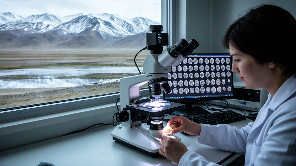
Tiny crustaceans no bigger than a grain of rice may soon reveal evolutionary secrets at a scale that was previously impossible, thanks to a new study that has brought automation to one of palaeontology’s most stubborn bottlenecks. Researchers led by Marlene Hoehle of the University of Greifswald have demonstrated that a high-throughput imaging pipeline originally developed for marine microfossils can reliably extract size and shape data from non-marine ostracods, the bivalved microcrustaceans whose calcified shells accumulate in lake sediments for millions of years. The work, published in the Journal of Micropalaeontology, tested the AutoMorph software package on two endemic ostracod species sampled from six lakes across the Tibetan Plateau, and the results suggest that the era of laboriously hand-digitising specimen outlines may finally be drawing to a close.
Ostracods occupy a special place in evolutionary and environmental research. They live in nearly every aquatic habitat on Earth, from the Arctic to the tropics, they produce calcified valves that fossilise readily, and they display remarkable taxonomic and morphological diversity. Because many species are endemic to individual lake systems, they serve as sensitive regional indicators of environmental conditions. Their short life cycles make them ideal for studying how populations respond to ecological pressures, and their long fossil records allow those responses to be traced across geological time. Yet despite these advantages, ostracods have remained underrepresented in quantitative morphological studies, largely because the methods available for measuring their form are slow, subjective, and difficult to scale.
The core problem lies in geometric morphometrics, the gold-standard framework for quantifying biological form. In the landmark-based approach, researchers place coordinate points on biologically or mathematically homologous features of each specimen, then use statistical procedures such as generalised Procrustes analysis to align all configurations for comparison. For ostracods this is particularly challenging, because non-marine taxa often have smooth, poorly ornamented valves that lack sufficient fixed points of reference. Researchers have therefore turned to outline-based methods, in which dozens of sliding semi-landmarks are placed along the valve margin. In previous studies, this meant manually positioning 68 to 93 points on every single valve, a process that limited datasets to a few hundred specimens from a handful of localities.
AutoMorph, a Python-based software package first developed for planktonic foraminifera, promised a way out. The pipeline extracts outlines of white objects against black backgrounds and can process thousands of specimens per day, enabling the construction of image datasets at scales previously unattainable in marine micropalaeontology. Those datasets have already transformed understanding of macroevolutionary and macroecological patterns in the global ocean. But continental settings present their own opportunities and challenges. Lakes are isolated, small-scale systems where adaptation, radiation, and evolutionary development can unfold in distinctive ways, and with roughly 117 million lakes larger than 0.2 hectares worldwide, the potential for comparative evolutionary studies is enormous.
The Tibetan Plateau, with nearly 10,000 water bodies spread across approximately 2.5 million square kilometres, offers an ideal natural laboratory. The team focused on two species endemic to the region’s large and deep lakes, Leucocythere dorsotuberosa and Leucocytherella sinensis, which dominate the ostracod faunas of these high-altitude ecosystems. Sediment surface samples collected during Sino-German field campaigns between 2008 and 2012 yielded 553 adult valves from lakes including Nam Co, Tangra Yumco, Chen Co, Xuru Co, Pumayum Co, and Npen Co. Specimens were photographed in bulk using the panorama function of a Keyence VHX 7000 digital microscope, which captured composite overview images of entire sub-datasets at 150-times magnification with extended depth of focus.
Adapting AutoMorph to ostracods required several technical modifications. The segmentation module, which detects and isolates individual objects within bulk images, was tuned with species-specific size ranges of 400 to 1,500 micrometres for L. sinensis and 700 to 2,000 micrometres for L. dorsotuberosa, with threshold values between 0.3 and 0.4 proving most effective. Because the picking trays used during imaging produced backgrounds that were insufficiently dark and sometimes marred by scratches, dust, or sediment particles, the team added a step that sets the image background to black before saving each segmented object. This seemingly small adjustment proved decisive: outline extraction subsequently succeeded for every single specimen, a 100 percent success rate that the authors note is a marked improvement over the original implementation.
The run2dmorph module then detected each valve outline through pixel brightness, measured parameters such as area, perimeter, and axis lengths, and placed 100 coordinate points along the outline. Because left and right valves are mirror images with different orientations, separate scripts were written for each side, defining the starting point at the maximum anterior margin and proceeding along the dorsal margin in the appropriate direction. A custom Python script identified the maximum rightmost and leftmost points as fixed landmarks, producing a final configuration of 102 coordinate points per specimen ready for generalised Procrustes analysis. Validation against manual measurements taken with the microscope’s integrated software showed average deviations of less than 2 percent, confirming that the automated measurements are as accurate as hand measurements while being vastly faster.
The biological results were equally encouraging. Principal component analyses of the shape data revealed clear separation between species and sexes in both left and right valves, with the first three principal components explaining between roughly 52 and 61 percent of shape variation for left valves and even more for right valves. Linear discriminant analysis based on the first 15 components cleanly distinguished species and sexes, and Procrustes analysis of variance confirmed that group means differed significantly. Interestingly, the analyses showed that male and female valves of the two species follow different size patterns: male L. dorsotuberosa valves are shorter and narrower than those of females, while male L. sinensis valves are larger. A sampling evaluation using the LaSEC function confirmed that around 80 coordinate points are sufficient to characterise shape variation, a density easily achieved by the automated workflow.
Beyond speed, automation tackles a subtler but equally important problem: observer bias. Studies of measurement error in geometric morphometrics have found that landmark placement can vary between and even within digitisers, and that combined error sources can exceed 30 percent of the total biological variation in a dataset, with inter-observer error the largest contributor. Automated coordinate generation eliminates this variability entirely, producing consistent output across specimens and substantially reducing subjectivity. The trade-off is that the automated approach demands well-preserved specimens, since even minimally damaged valves produce distorted outlines that manual methods might accommodate. The authors argue that this constraint is outweighed by the ability to process far larger numbers of intact specimens, and that damaged individuals can be flagged through outlier detection before analysis.
The implications reach well beyond the Tibetan Plateau. Automated geometric morphometrics could transform ostracods into model organisms for studying evolutionary and ecological dynamics across spatial and temporal scales that were previously impractical, from comparing populations across dozens of lakes to tracking morphological change through sediment cores such as those expected from the International Continental Scientific Drilling Program’s NamCore project at Nam Co. The authors also point toward shared image and morphometric databases that would support machine learning applications, which are already well developed for foraminifera and diatoms but remain in their infancy for ostracods. Integrating shape data with environmental variables and, where possible, genetic information could finally disentangle the drivers of morphological variation, whether genetic, environmental, or the result of phenotypic plasticity. What once took weeks or months of painstaking manual work can now be accomplished in hours, opening a window onto the hidden geometry of evolution in some of Earth’s most isolated aquatic ecosystems.
Subject of Research: Automated geometric morphometric analysis of non-marine ostracod valves from Tibetan Plateau lakes
Article Title: Testing the applicability of automated size and shape analyses in non-marine ostracods – a case study from the Tibetan Plateau
Article References: Hoehle, M., Haberzettl, T., Frenzel, P., Schwalb, A., Wang, J., Zhu, L., & Wrozyna, C. (2026). Testing the applicability of automated size and shape analyses in non-marine ostracods – a case study from the Tibetan Plateau. Journal of Micropalaeontology, 45(1), 359-375. https://doi.org/10.5194/jm-45-359-2026
Image Credits: AI Generated
Keywords: ostracods, geometric morphometrics, AutoMorph, Tibetan Plateau, automation, shape analysis, micropalaeontology, evolutionary biology, lake sediments, image processing, sexual dimorphism, endemism
News Source: Violet Maxwell. (October 9, 2026). Automated Imaging Pipeline Cracks the Bottleneck in Ostracod Shape Analysis on the Tibetan Plateau. Scienmag.