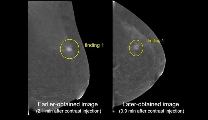
Honolulu, HI | April 19, 2023—Findings from a Scientific Online Poster presented during the 2023 ARRS Annual Meeting at the Hawaiian Convention Center suggest there is institutional variability in both projection order and image acquisition timing for contrast-enhanced mammography (CEM) protocol, with a previous systematic review revealing at least 7 different combinations in projection order.

Credit: ARRS
Honolulu, HI | April 19, 2023—Findings from a Scientific Online Poster presented during the 2023 ARRS Annual Meeting at the Hawaiian Convention Center suggest there is institutional variability in both projection order and image acquisition timing for contrast-enhanced mammography (CEM) protocol, with a previous systematic review revealing at least 7 different combinations in projection order.
“Our study demonstrates that earlier-obtained recombined imaging is significantly preferred in cancer lesion characterization, with a few instances demonstrating that biopsy-proven lesions may appear more conspicuously on earlier-obtained imaging (e.g., mass versus non-mass enhancement),” said Dennis Dwan, MD, of Beth Israel Deaconess Medical Center in Boston, MA. Also noting a preference trend for craniocaudal (CC) over mediolateral oblique (MLO) projection, “given better characterization on earlier imaging,” Dr. Dwan added, “CEM protocols should consider prioritizing imaging the side of pathology.”
In Dwan et al.’s study, two groups of consecutive CEM cases were performed on patients prior to cancer diagnosis: 40 cases, from 2016 to 2018, acquired with CC views performed first; 38 cases, from 2019 to 2020, acquired with MLO views performed first. The side of pathology was initially imaged in either the CC or MLO projection (earlier-obtained imaging), followed by the contralateral side in the same projection. Then, imaging was repeated for the other projection (later-obtained imaging). Five readers evaluated cases for cancer visibility, confidence margin, and cancer conspicuity against background parenchymal enhancement (BPE). Additional analysis compared earlier- and later-obtained images, as well as CC compared to MLO projection.
In this ARRS Annual Meeting Online Poster, 78 female patients were included (mean age, 58 years). Ultimately, no significant difference was shown in menopausal status, breast density, or BPE between group 1 and 2. Mean acquisition time of the earlier- and later-obtained imaging was 2.5 minutes and 4.75 minutes, respectively. In 35/390 (9%) of instances, an individual reader changed the reported lesion type between earlier and later imaging, with most instances (n = 28/35, 80%) reflecting a situation where a finding was downgraded to a less conspicuous finding on later-obtained imaging.
Overall, reader preference was for earlier-obtained images when assessing visibility of cancer, confidence in margins, and conspicuity of lesion against BPE (p < 0.001). Among all readers, there was preference for CC views over MLO projection when evaluating lesion conspicuity against BPE (p = 0.045) and no significant preference for the evaluation of lesion visibility (p = 0.078) or confidence in margins (p = 0.35).
North America’s first radiological society, the American Roentgen Ray Society (ARRS) remains dedicated to the advancement of medicine through the profession of medical imaging and its allied sciences. An international forum for progress in radiology since the discovery of the x-ray, ARRS maintains its mission of improving health through a community committed to advancing knowledge and skills with the world’s longest continuously published radiology journal—American Journal of Roentgenology—the ARRS Annual Meeting, InPractice magazine, topical symposia, myriad multimedia educational materials, as well as awarding scholarships via The Roentgen Fund®.
MEDIA CONTACT:
Logan K. Young, PIO
44211 Slatestone Court
Leesburg, VA 20176
Method of Research
Imaging analysis
Subject of Research
People
COI Statement
N/A


