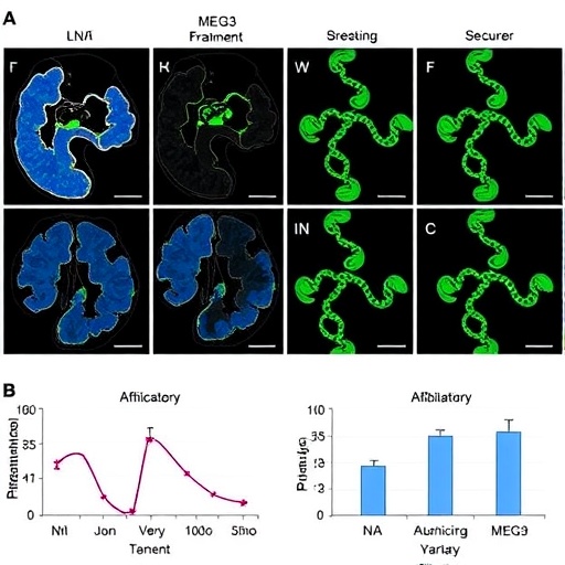
Parkinson’s disease may be leaving behind a molecular trail far more intricate than previously understood. A new study published in npj Parkinson’s Disease reports that small RNA fragments derived from alanine transfer RNA interact with a long splice variant of the long non-coding RNA MEG3, and that these molecular relationships change according to the tissue examined, the cellular compartment involved and the severity of the disease. The findings add another layer to the biology of Parkinson’s, a neurodegenerative disorder traditionally associated with the progressive loss of dopamine-producing neurons in a brain region called the substantia nigra. Rather than presenting disease as a single, uniform molecular process, the research points toward a shifting network of RNA signals that may behave differently in different biological environments.
The molecules at the center of the study belong to two classes of RNA that do not produce proteins in the conventional way. Transfer RNAs, or tRNAs, are best known for delivering amino acids to the ribosome during protein synthesis. Alanine tRNAs help carry the amino acid alanine, but they can also be cut into shorter molecules known as tRNA-derived fragments, or tRFs. These fragments are not simply cellular debris. Depending on their sequence, length and chemical modifications, they may influence messenger RNA stability, protein production, stress responses and communication between cells. Their abundance can change during inflammation, metabolic disruption and neurodegeneration, making them potential indicators of cellular stress as well as possible regulators of disease biology.
The second component, MEG3, is a long non-coding RNA, meaning it is transcribed from DNA but does not primarily serve as a template for making a protein. Long non-coding RNAs can act as molecular scaffolds, guides or competing binding partners, bringing regulatory proteins and nucleic acids into proximity. They may influence gene expression in the nucleus, affect RNA processing or alter the fate of molecules in the cytoplasm. The study focuses specifically on a long splice variant of MEG3. Splicing allows cells to assemble different RNA versions from the same gene, and these variants can possess distinct structures, cellular locations and molecular partners. A splice variant is therefore not merely a longer copy; it may represent a functionally different regulatory molecule.
Zorbaz, Bennett, Treidel and colleagues examined the relationship between alanine tRNA fragments and this MEG3 long splice variant in the context of Parkinson’s disease. According to the study’s title, the interaction does not remain constant. Instead, it shows tissue-dependent changes, indicating that the molecular partnership may differ between biological tissues. It also varies by cellular compartment, suggesting that the location of the RNA molecules—such as the nucleus, cytoplasm or other intracellular fractions—matters to their behavior. Finally, the interaction changes with disease severity, raising the possibility that the RNA network is dynamically remodeled as Parkinson’s progresses rather than remaining fixed from its earliest stages.
That distinction is important because RNA molecules are highly sensitive to their surroundings. The same sequence can have different effects depending on whether it is inside the nucleus, where gene regulation and RNA processing occur, or in the cytoplasm, where messenger RNA translation and degradation are more prominent. RNA abundance alone may therefore provide an incomplete picture. Two patients could show similar overall levels of a particular tRNA fragment, while the fragment occupies different compartments or is associated with different molecular partners. By examining location and interaction together, researchers can begin to distinguish between an RNA molecule that is actively participating in disease mechanisms and one that is merely accumulating as a consequence of cellular damage.
The findings also reinforce the emerging view that Parkinson’s disease involves extensive disruption of RNA biology. Neurons depend on carefully timed transport, translation and degradation of RNA to maintain long axons and specialized synaptic connections. They must continually adjust protein production in response to electrical activity, energy availability and environmental stress. Misfolded proteins, mitochondrial dysfunction, impaired waste clearance and neuroinflammation can all disturb this balance. Small tRNA fragments and long non-coding RNAs may become part of the cell’s response, either helping it adapt or contributing to dysfunction when regulation breaks down. Their interaction could influence which messenger RNAs are translated, how long those messages persist or how cells communicate stress signals.
The severity-dependent pattern is particularly intriguing because it suggests that the alanine tRNA fragment–MEG3 relationship could reflect disease stage. However, a changing molecular signature does not automatically prove that it drives the progression of Parkinson’s. It may be a cause, a consequence or part of a feedback loop in which neuronal injury alters RNA regulation and altered RNA regulation intensifies cellular vulnerability. Establishing direction will require longitudinal studies that track individuals over time, as well as experimental work in neurons and disease models. Researchers will also need to determine whether the molecular pattern is specific to Parkinson’s disease or appears more broadly in aging, inflammation and other neurodegenerative conditions.
If validated, the RNA interaction could eventually have value as a biomarker. Biomarkers are measurable biological features that help detect disease, estimate prognosis or monitor a response to treatment. RNA fragments are attractive candidates because they can sometimes be detected in accessible fluids, including blood or cerebrospinal fluid, and because their levels may shift before extensive tissue damage becomes clinically obvious. Yet the study’s emphasis on tissue and cellular compartment is a reminder that translation into a clinical test will not be straightforward. A signal measured in blood may not directly mirror the molecular events occurring inside vulnerable neurons. Accurate interpretation may require panels of RNA molecules, information about their chemical modifications and methods capable of distinguishing free fragments from fragments carried inside extracellular vesicles.
The work may also open a path toward RNA-based intervention. In principle, synthetic oligonucleotides could be designed to block an unwanted interaction, stabilize a protective RNA structure or redirect a fragment toward a particular target. Similar strategies are being explored in other neurological and genetic diseases, although delivery into the human brain remains a major challenge. RNA medicines must reach the correct cells, enter them efficiently and avoid provoking harmful immune reactions. The complex, compartment-specific nature of the interaction described in this study makes precision especially important: changing the abundance of an RNA molecule throughout the body could produce effects very different from selectively altering its activity in affected neurons.
For now, the most significant message is conceptual. Parkinson’s disease appears increasingly less like a single pathway that fails and more like a moving molecular ecosystem in which regulatory RNAs, proteins, organelles and cell types influence one another over time. The discovery of tissue-, compartment- and severity-dependent changes involving alanine tRNA fragments and a MEG3 splice variant adds detail to that ecosystem and highlights why molecular context matters. As scientists continue mapping these RNA networks, they may uncover signatures capable of revealing disease biology earlier, separating subtypes more accurately and identifying treatments matched to an individual’s stage and molecular profile. The study does not yet offer a new therapy, but it brings researchers closer to understanding how the RNA language of stressed neurons changes as Parkinson’s disease advances.
Subject of Research: Alanine tRNA fragments and their interaction with a long splice variant of the MEG3 long non-coding RNA in Parkinson’s disease.
Article Title: Alanine tRNA fragments interacting with MEG3 long splice variant show tissue-, cellular compartment- and disease severity-dependent changes in Parkinson’s disease.
Article References: Zorbaz, T., Bennett, E.R., Treidel, S.V. et al. “Alanine tRNA fragments interacting with MEG3 long splice variant show tissue-, cellular compartment- and disease severity-dependent changes in Parkinson’s disease.” npj Parkinson’s Disease (2026). https://doi.org/10.1038/s41531-026-01541-2
Image Credits: AI Generated
DOI: 10.1038/s41531-026-01541-2
Keywords: Parkinson’s disease, alanine tRNA fragments, tRNA-derived fragments, MEG3, long non-coding RNA, RNA interactions, RNA biology, neurodegeneration, biomarkers, disease severity
Tags: impact of tRFs on neuronal health and diseaselong non-coding RNA MEG3MEG3 long non-coding RNA variants in Parkinson’snon-coding RNAs as potential biomarkers for Parkinson’s severityParkinson’s disease molecular biomarkersRNA signaling networks in Parkinson’s disease progressionrole of alanine tRNA fragments in neurodegenerative disorderstissue compartment differences in Parkinson’s molecular biologytissue-specific RNA interactions in Parkinson’stRNA-derived fragments in neurodegeneration