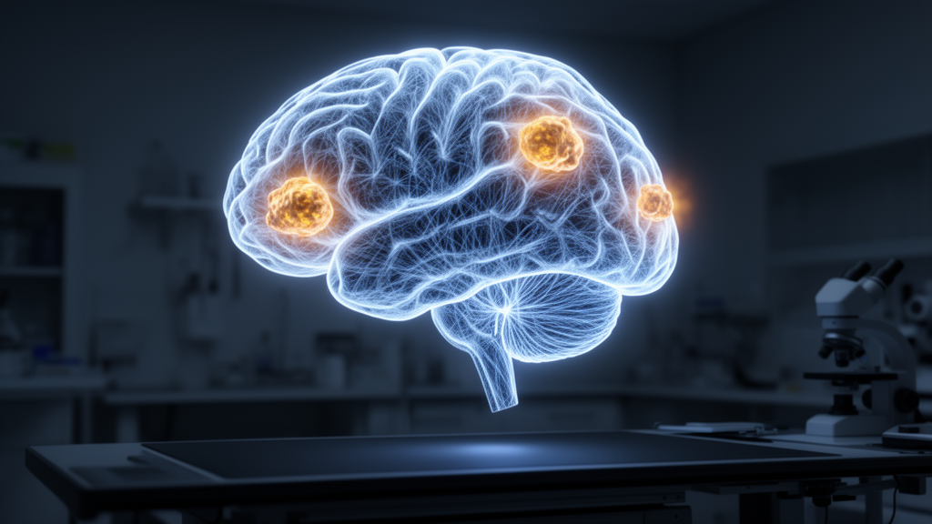
Artificial intelligence systems that read brain scans are often celebrated for their accuracy, but a new study argues that the more important question is not whether the model is right — it is whether the model knows when it might be wrong. Researchers at Binghamton University have developed SYNAPSE-X, a calibration-aware Vision Transformer framework that classifies brain tumor MRI scans into four categories while explicitly measuring how trustworthy its own confidence scores are. The work, published in Neural Computing and Applications, reframes medical image classification as a reliability problem rather than a pure prediction contest, and its results suggest that a model which can defer uncertain cases to human experts may be far more useful in practice than one that simply reports a single headline accuracy figure.
The framework, developed by Sadaf Tabatabaee and Mohammad T. Khasawneh, was trained to distinguish glioma, meningioma, pituitary tumor, and no-tumor cases from contrast-enhanced T1-weighted MRI images. The corrected experimental cohort contained 7,023 images, split into 5,712 training images and 1,311 held-out test images, with data augmentation applied only to the training subset to reduce the risk of information leaking between splits. On the untouched test set, SYNAPSE-X achieved 98.41 percent accuracy, 98.2 percent macro-F1, and 98.4 percent weighted-F1 — figures comparable to the best convolutional and transformer-based pipelines previously reported on similar public four-class brain MRI datasets.
What sets the study apart is what happened next. The researchers point out that a model can be highly accurate on average yet dangerously overconfident in its errors, a phenomenon documented since Guo and colleagues showed in 2017 that modern neural networks are frequently miscalibrated. Calibration means that when a model says it is 90 percent sure, it should be right roughly 90 percent of the time on similar predictions. To measure this, the team used three complementary metrics: expected calibration error, which quantifies the gap between stated confidence and observed accuracy across confidence bins; the Brier score, a classical measure of how close predicted probabilities are to actual outcomes; and negative log-likelihood, which harshly penalizes confident mistakes.
Before any correction, SYNAPSE-X showed moderate calibration error despite its stellar accuracy. The team then applied temperature scaling, a simple post hoc technique that rescales the model’s raw output logits with a single learned parameter before the softmax function converts them into probabilities. Crucially, the temperature parameter was fitted only on a validation subset, never on the test data or the external cohort, preventing calibration leakage. The result was striking: expected calibration error fell from 0.041 to 0.018, a relative reduction of roughly 56 percent, while the Brier score dropped from 0.036 to 0.021 and negative log-likelihood fell from 0.092 to 0.061. Because temperature scaling only reshapes probability values and never changes which class the model picks, classification accuracy remained exactly the same — but the confidence numbers became genuinely meaningful.
With calibrated confidence in hand, the researchers implemented the framework’s most distinctive feature: selective referral. Instead of forcing every scan through the same automated decision pathway, predictions whose calibrated confidence fell below a threshold of 0.85 were flagged for expert review, while high-confidence predictions were retained for decision-support reporting. At this operating point, 93.3 percent of internal test cases were retained and 6.7 percent were referred to human specialists. Among the retained cases, accuracy rose to 99.12 percent, pushing retained-case risk below one percent. Sensitivity analyses across thresholds from 0.70 to 0.95 traced out the expected risk-coverage trade-off: more conservative thresholds reduced risk among retained cases but increased the referral burden on clinicians.
The error profile analysis added a clinically reassuring pattern. Referred and misclassified cases were not scattered randomly across the dataset; they clustered in categories where visual overlap is expected, particularly between glioma and meningioma, followed by meningioma and pituitary tumor. Meningioma showed the highest referral tendency, consistent with the fact that meningiomas can share enhancement patterns and boundary characteristics with other tumor types depending on location and imaging plane. No-tumor cases, by contrast, carried the lowest referral burden and the strongest high-confidence behavior. This suggests the model’s uncertainty is concentrated exactly where a radiologist would expect diagnostic difficulty — a property that makes confidence-based triage genuinely informative rather than arbitrary.
Perhaps the most demanding test came from outside the development data entirely. The trained model was applied, without any retraining, fine-tuning, architecture modification, external recalibration, or threshold re-selection, to an independent external brain MRI cohort of 3,264 images. Under this direct-transfer regime, SYNAPSE-X achieved 97.86 percent accuracy, 97.7 percent macro-F1, and 97.9 percent weighted-F1, with an external expected calibration error of just 0.026. At the same 0.85 referral threshold, the external referral rate was 8.4 percent and retained-case accuracy remained high at 98.74 percent. The modest performance drop relative to internal testing is exactly what dataset shift would predict, and the fact that calibration and referral behavior survived the transfer intact strengthens the case that the framework’s reliability signals are robust rather than artifacts of a single dataset.
The authors are careful to position the work honestly. Both the primary and external cohorts are public image-level datasets lacking complete patient-level identifiers, scanner metadata, and institutional acquisition information, which means patient-level independence could not be verified. The study is therefore framed as research-stage reliability assessment rather than clinical validation, and the external testing is described as direct-transfer evaluation rather than proof of deployment readiness. This cautious framing aligns with reporting guidelines such as CLAIM and TRIPOD+AI, and with a broader literature showing that radiology AI models often lose performance across hospital sites, sometimes because they learn site-specific acquisition signatures instead of genuine disease features. Distributional shift, prior work has shown, can degrade the reliability of confidence values themselves, not just accuracy — which is precisely why calibration-aware external testing matters.
Technically, the underlying classifier is a compact Vision Transformer in the tradition introduced by Dosovitskiy and colleagues, in which each 224-by-224 pixel MRI image is divided into non-overlapping 16-by-16 patches, producing 196 patch embeddings. Positional embeddings are added, and the sequence passes through transformer encoder blocks built on multi-head self-attention, residual connections, layer normalization, and feed-forward layers before a four-unit softmax head produces class probabilities. This architecture allows the model to capture not only local texture but also lesion shape and spatial context, which matters when tumor discrimination depends on relationships across distant image regions. Training used categorical cross-entropy loss, AdamW optimization with weight decay, dropout, and training-only augmentation, with checkpoint selection based on validation loss and macro-F1.
The broader significance of SYNAPSE-X lies in its conceptual shift. Conventional classification studies report one aggregate accuracy number, implicitly treating every prediction as equally dependable. By jointly evaluating accuracy, probability calibration, confidence-stratified correctness, risk-coverage behavior, and external transfer stability, the framework converts brain tumor MRI classification into a reliability-aware selective-referral problem in which the AI acts as a triage-aware assistant rather than an infallible oracle. The authors suggest that future work should move to patient-level, multi-institutional datasets with scanner diversity, prospective evaluation, and radiologist-in-the-loop studies to determine whether calibrated selective referral can support safe clinical workflows. For now, the study offers a compelling demonstration that the next generation of medical AI may be judged not by how often it is right, but by how honestly it communicates the moments when it is not sure.
Subject of Research: Calibration-aware Vision Transformer classification of brain tumor MRI with uncertainty-guided selective referral
Article Title: SYNAPSE-X: a calibration-aware vision transformer framework for selective referral in multi-class brain tumor MRI classification
Article References: Tabatabaee, S., & Khasawneh, M. T. (2026). SYNAPSE-X: a calibration-aware vision transformer framework for selective referral in multi-class brain tumor MRI classification. Neural Computing and Applications, 38(20), Article 813. https://doi.org/10.1007/s00521-026-12608-9
Image Credits: AI Generated
DOI: 10.1007/s00521-026-12608-9
Keywords: brain tumor, MRI, Vision Transformer, deep learning, calibration, temperature scaling, selective referral, uncertainty quantification, medical imaging, risk-coverage analysis, external validation, decision support
News Source: Nathaniel Bowman. (October 17, 2026). AI That Knows When It’s Unsure: New Brain Tumor Model Flags Its Own Doubts. Scienmag.