Periodontal disease is one of the most common chronic conditions on the planet, and one of its most telling signs hides in plain sight on a routine dental X-ray: the slow, silent erosion of the bone that anchors teeth in the jaw. Reading that erosion, however, is as much an art as a science. Dentists squinting at panoramic radiographs must judge how far the bone has receded relative to the tooth root, a judgment that varies from one observer to the next and can shift even for the same observer on different days. A new study published in BMC Medical Imaging suggests that artificial intelligence can shoulder part of that burden, and that the secret to doing it well is not just looking at the image but also reading the chart beside it.
Researchers Adnan Salman of the Computer Science Department and Ibtehal Raghib Bishtawi of the Faculty of Dentistry and Dental Surgery, both at An-Najah National University in Palestine, built a multimodal deep learning framework that fuses two very different kinds of information: the pixel-rich world of panoramic radiographs and the structured metadata that accompanies them, such as patient age and other clinical variables recorded in the dataset. Their target was the automated classification of periodontal bone resorption severity using the Bone Resorption Age Ratio, or BRAR, a measure that relates the degree of bone loss to the patient’s age. The work, published open access on 13 October 2026, arrives at a moment when dental practices are drowning in imaging data and clinicians are increasingly open to algorithmic second opinions.
The technical architecture behind the study is a study in pragmatic engineering. For the imaging pathway, the team employed DenseNet-121, a convolutional neural network celebrated for its densely connected layers, in which each layer receives feature maps from all preceding layers. That design encourages feature reuse, keeps parameter counts modest, and has made DenseNet variants a workhorse of medical image analysis. The metadata pathway was handled by a multilayer perceptron, a classical feedforward neural network that transforms tabular inputs into a compact numeric representation. The two streams were then combined through feature-level concatenation, meaning the learned image features and the learned metadata features were joined end to end into a single vector before the final classification layers made their verdict. The combined network was refined with progressive fine-tuning, a training strategy in which parts of the network are unfrozen and retrained in stages, allowing the model to adapt pretrained image representations to the dental domain without erasing what they already know.
The evidence base consisted of 988 annotated panoramic radiographs drawn from a publicly available dataset, collected and de-identified under ethical approval from the Institutional Ethics Committee of Shanghai Stomatological Hospital. To guard against the vagaries of a single lucky train-test split, a notorious pitfall in machine learning research, the authors evaluated image-only, metadata-only, and multimodal models across 20 repeated stratified random splits. This repeated-split design yields a distribution of performance estimates rather than a single number, and it enabled rigorous paired statistical comparison. The team assessed accuracy, macro F1-score, macro area under the receiver operating characteristic curve, and average precision, along with class-specific metrics that reveal how the model treats each severity level. Differences in three primary outcomes, namely macro F1, macro AUC-ROC, and recall for the most severe category, were tested with the Wilcoxon signed-rank test and corrected for multiple comparisons using the Holm-Bonferroni procedure, a combination that raises the bar for claims of statistical significance.
The headline numbers tell a story of meaningful, if imperfect, progress. The multimodal model achieved an accuracy of 0.67 plus or minus 0.03, a macro F1-score of 0.65 plus or minus 0.03, and a macro AUC-ROC of 0.81 plus or minus 0.03. Perhaps most importantly for clinical triage, its recall on Level 3, the severe class, reached 0.60 plus or minus 0.07, meaning it caught roughly six in ten of the worst cases. Those figures may look modest next to the near-perfect scores that dominate headlines in other corners of AI medicine, but severity grading from a single two-dimensional projection of a three-dimensional jaw is a genuinely hard problem, and the comparisons are what give the results their weight.
And the comparisons were decisive. The multimodal model significantly outperformed the image-only model on macro F1 and macro AUC-ROC, both with Holm-adjusted p-values below 0.001, and on Level 3 recall with a p-value of 0.036. It also beat the metadata-only model on all three primary outcomes with p-values below 0.001. The metadata-only model deserves particular attention, because it exposed the danger of trusting demographics alone: it identified only 0.14 plus or minus 0.07 of severe cases, a failure rate that would be clinically unacceptable. In other words, neither the picture nor the chart is sufficient on its own, but together they are substantially better than either alone.
One obvious objection demanded testing: perhaps the metadata contribution was nothing more than the patient’s age, which is already baked into the BRAR definition. If so, the multimodal gains would be a kind of circular bookkeeping rather than genuine added insight. The authors therefore ran a four-way metadata ablation, systematically removing or isolating inputs to measure each one’s contribution. The result was clarifying. Neither age alone nor the five remaining non-age structured variables alone significantly improved macro F1 or Level 3 recall over the image-only model, whereas the full metadata set did. The improvement, in other words, emerges from the complementary contributions of age and the other structured variables acting together, a finding that speaks to the subtle ways heterogeneous data sources can reinforce one another when a neural network learns to weigh them jointly.
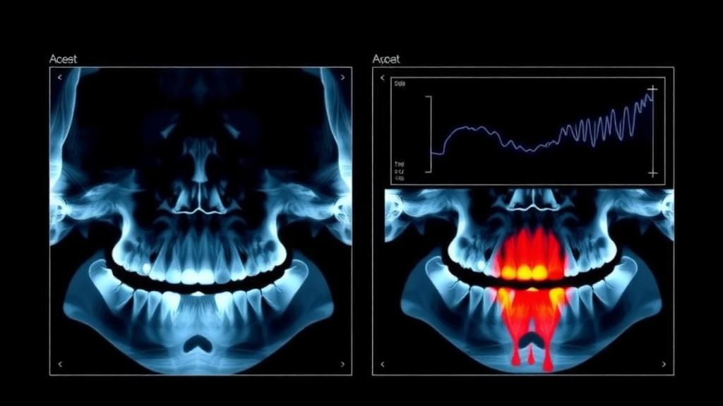
The study went further than aggregate statistics. The researchers generated Gradient-weighted Class Activation Mapping, or Grad-CAM, visualizations, a technique that highlights the image regions most influential in the network’s decision, offering a qualitative window into whether the model attends to clinically plausible areas such as the alveolar bone crests. They also explored a probability-level ensemble, combining model outputs rather than features, and reported those results as exploratory. Most intriguingly, they conducted a targeted expert error analysis on 88 purposively selected cases, asking a dentist to independently grade a sample that included cases the model got right and cases it got wrong. The pattern was striking: dentist-annotation agreement was considerably higher among model-correct cases, with a kappa of 0.585 and a weighted kappa of 0.650, than among model-misclassified cases, where kappa fell to 0.208 and weighted kappa to 0.248. Kappa statistics quantify agreement beyond chance, and values in the 0.2 range indicate only fair agreement at best.
That finding hints at an uncomfortable but important possibility: some of the model’s errors may overlap with zones of genuine human disagreement, where the severity label itself is ambiguous. Yet the authors are careful not to overclaim. Because the expert analysis involved a single dentist examining a purposively selected subset rather than a random sample, it cannot establish annotation uncertainty as the cause of the model’s mistakes. This restraint is characteristic of the paper’s overall tone, which presents the multimodal gains as robust while flagging the limits of what the evidence can support. It is a refreshing contrast to the breathless framing that often surrounds medical AI, and it gives clinicians a realistic picture of what the system can and cannot yet do.
The road ahead, as the authors outline it, involves three main thrusts: external validation on datasets beyond the one used here, automated localization of bone resorption on the radiograph rather than whole-image classification, and a shift toward direct radiographic estimation of bone resorption followed by age normalization, which could decouple the measurement from the label scheme. The study received no specific grant funding, and the authors declare no competing interests. What it delivers today is a proof of principle with a rigorous statistical backbone: that the humble dental chart, the age and structured details clinicians record every day, carries signal that a well-designed neural network can exploit to judge jawbone loss more reliably. As panoramic radiographs remain among the most widely acquired images in all of medicine, even modest, verified gains in automated severity assessment could ripple through screening programs, dental schools, and busy clinics, wherever the quiet erosion of bone needs a second, tireless pair of eyes.
Subject of Research: Automated assessment of periodontal bone resorption severity using multimodal deep learning on panoramic radiographs and structured metadata
Article Title: Multimodal deep learning for automated assessment of periodontal bone resorption severity using panoramic radiographs and structured metadata
Article References: Multimodal deep learning for automated assessment of periodontal bone resorption severity using panoramic radiographs and structured metadata. (n.d.). https://doi.org/10.1186/s12880-026-02928-6
Image Credits: AI Generated
DOI: 10.1186/s12880-026-02928-6
Keywords: multimodal deep learning, panoramic radiographs, periodontal disease, bone resorption, BRAR, DenseNet-121, medical image analysis, Grad-CAM, dental radiology, machine learning, severity classification, metadata fusion
News Source: Blake Davidson. (October 13, 2026). AI Reads Dental X-Rays and Patient Data Together to Gauge Gum Disease Severity. Scienmag.