For women with breast cancer that has spread to the lymph nodes under the arm, one of the most consequential questions after months of chemotherapy is deceptively simple: did the drugs actually wipe out every cancer cell in those nodes? The answer, today, can only be confirmed by surgery. A team of radiologists in China now reports that an artificial intelligence model, reading standard contrast-enhanced MRI scans, can make that prediction noninvasively with striking accuracy — and in doing so, could spare thousands of patients an operation they may not need.
The study, published in BMC Medical Imaging, was led by Jun Liao, Mengting Xu and Ruimin Li, with corresponding author Hongna Tan, at the People’s Hospital of Zhengzhou University and Henan Provincial People’s Hospital, together with collaborators at Fudan University Shanghai Cancer Center. The researchers set out to tackle a long-standing blind spot in breast cancer care: the axillary lymph nodes, the first and most common destination for breast cancer cells leaving the tumor, are notoriously difficult to assess with imaging alone. Yet knowing whether chemotherapy has eradicated the disease there — a state clinicians call pathological complete response, or pCR — is exactly the information that determines whether a patient can safely avoid a full axillary lymph node dissection, an operation that removes most of the lymph nodes under the arm and is a leading cause of lifelong arm swelling, known as lymphedema.
Neoadjuvant chemotherapy — drugs given before surgery to shrink the tumor — has transformed breast cancer treatment over the past two decades. When it works completely, obliterating both the breast tumor and any nodal metastases, patients with node-positive disease can often be managed with far less invasive surgery. The problem is verification. Sentinel lymph node biopsy, the standard minimally invasive check, can miss residual disease, and many centers still default to full axillary dissection for patients who had node-positive disease at diagnosis, precisely because they cannot confidently rule out remaining cancer. The result, the authors note, is a substantial rate of unnecessary operations: in their retrospective simulation, a conventional approach would have led to axillary lymph node dissection in roughly 40.7 percent of patients who did not actually need it.
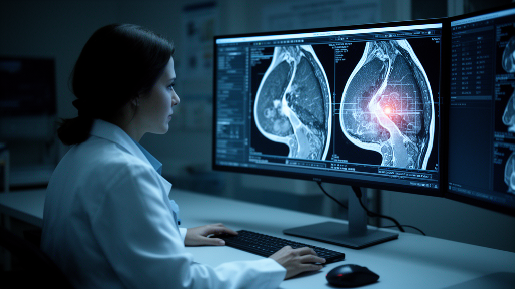
The researchers’ answer was to build a deep learning system that looks at more of the picture than previous attempts. Their model ingests dynamic contrast-enhanced MRI — the workhorse breast imaging technique in which a gadolinium contrast agent reveals how blood flows through tissue over time — from multiple regions simultaneously: the primary tumor in the breast and the involved axillary lymph nodes. Crucially, they compared two architectural philosophies. A 2D model processes each MRI slice in isolation, treating every cross-section as a separate image. A 2.5D model stacks adjacent slices together, feeding the network a thin slab of tissue at a time so it can perceive features that only make sense in three dimensions — the way contrast washes in and out of a lesion, the shape of a node’s cortex, the texture of its interior across depth.
The difference proved decisive. In a validation set of 91 patients held out from training, the 2.5D tumor-only model achieved an area under the receiver operating characteristic curve, or AUC, of 0.797, comfortably ahead of the 0.706 posted by its 2D counterpart. When the model was given both the tumor and the lymph nodes as separate input channels, performance climbed further: the 2.5D tumor-plus-node model reached an AUC of 0.834, versus 0.815 for the 2D version. The AUC is a standard measure of diagnostic discrimination — a value of 1.0 means perfect separation between patients who achieved pCR and those who did not, while 0.5 is equivalent to a coin flip. Values in the low 0.80s represent clinically meaningful predictive power.
The team then went a step further, fusing the imaging-derived features with clinicopathological data — estrogen receptor status, HER2 status and tumor grade, the three molecular and cellular characteristics that emerged from their statistical analysis as independent predictors of nodal response. The combined model, which the researchers call the 2.5D tumor–ALN–clinicopathologic model, delivered the best performance of anything they tested: an AUC of 0.861 in the validation cohort. Feature selection was rigorous, using the Mann–Whitney U test, Spearman correlation, a max-relevance and min-redundancy filter, and the least absolute shrinkage and selection operator, a technique known as LASSO that prunes variables down to the most informative subset while guarding against overfitting.
Perhaps the most striking result is not the raw accuracy but what it could mean in practice. The researchers ran a retrospective simulation: if the model’s predictions had been used to guide axillary management, the estimated rate of unnecessary axillary lymph node dissection would have fallen from 40.7 percent to 8.8 percent, while the estimated overall benefit rate — patients correctly spared an operation or correctly flagged for one — would have risen from 59.3 percent to 74.7 percent. Those are modeled estimates, not outcomes from a prospective trial, and the authors are careful to frame them as such. But the direction of the effect is hard to ignore: a tool that reads images already being acquired in routine care could, in principle, prevent the majority of avoidable node dissections.
The study cohort comprised 305 patients with invasive breast cancer and confirmed metastasis to the ipsilateral axillary lymph nodes, randomly split into a training set of 214 and a validation set of 91. Among them, 46.6 percent — 142 patients — achieved pathological complete response in the nodes after neoadjuvant chemotherapy, a proportion consistent with modern regimens in biologically favorable disease. The retrospective design, approved by the ethics committee of Henan Provincial People’s Hospital under Declaration of Helsinki protocols with informed consent waived, means the model has not yet been tested prospectively on patients whose treatment decisions were actually influenced by its output. That step — a validation study where the algorithm’s prediction is available to surgeons before they operate — is the necessary next chapter.
The work also reflects a broader shift in oncology imaging. Rather than asking a radiologist to eyeball whether a lymph node looks smaller or less bright on a follow-up scan, deep learning models can extract quantitative signatures of treatment response — patterns of enhancement kinetics, texture heterogeneity, spatial relationships between tumor and node — that are invisible or ambiguous to the human eye. Multichannel architectures, which feed different anatomical regions or image sequences into the same network as parallel inputs, are particularly well suited to this problem because nodal response and primary tumor response are related but not identical phenomena; a patient’s breast tumor may melt away while microscopic disease persists in the nodes, and vice versa. By modeling both regions jointly, the network can learn the interplay that single-region models miss.
For patients, the near-term significance is straightforward. Axillary lymph node dissection carries real, lasting costs: lymphedema, numbness, restricted shoulder mobility and increased infection risk in the affected arm. Any tool that can reliably identify which node-positive patients have achieved complete nodal clearance — without putting them under the knife to find out — would change the calculus of post-chemotherapy surgery. The 2.5D multichannel model described here is not yet that tool, but it is a credible step toward it, built on imaging that most breast cancer patients already receive as part of standard care. If prospective validation confirms the retrospective numbers, the algorithm could move from research code to clinical decision support, giving surgeons the confidence to offer less invasive nodal management to the patients who no longer need the more radical option.
Subject of Research: Deep learning prediction of axillary pathological complete response after neoadjuvant chemotherapy in breast cancer using multiregional DCE-MRI
Article Title: Multichannel deep learning using multiregional DCE-MRI to predict axillary pathological complete response after neoadjuvant chemotherapy in breast cancer
Article References: Liao, J., Xu, M., Li, R., Li, S., Cao, Z., Wang, D., Li, Y., Wang, M., & Tan, H. (2026). Multichannel deep learning using multiregional DCE-MRI to predict axillary pathological complete response after neoadjuvant chemotherapy in breast cancer. BMC Medical Imaging. https://doi.org/10.1186/s12880-026-02736-y
Image Credits: AI Generated
DOI: 10.1186/s12880-026-02736-y
Keywords: breast cancer, deep learning, DCE-MRI, axillary lymph nodes, pathological complete response, neoadjuvant chemotherapy, artificial intelligence, radiology, lymph node dissection, medical imaging, oncology, predictive modeling
News Source: Nathaniel Bowman. (October 17, 2026). AI Reads Breast MRI to Predict Whether Chemotherapy Has Cleared the Lymph Nodes. Scienmag.