When patients receive the contrast agent gadoxetic acid, healthy hepatocytes take up the molecule, and the signal intensities recorded across successive scan phases encode perfusion and hepatocellular function. Yet turning those raw signal values into meaningful indices has demanded painstaking manual work — radiologists placing small circular regions of interest on the liver, spleen, and back muscles, phase by phase, while avoiding vessels, ducts, and tumors. A new retrospective study from a tertiary academic hepatobiliary referral center, published in European Radiology Experimental, reports a deep learning pipeline that removes nearly all of that manual burden, quantifying five signal intensity-derived liver indices across the whole liver and every contrast phase without ever aligning one phase to another.
The research team, led by Felix Barajas Ordonez and Sven Nebelung, screened 9,013 liver MRI examinations performed between January 2016 and May 2025 and identified 3,112 that used gadoxetic acid. After excluding scans with incomplete dynamic phase coverage, severe artifacts, or non-fat-suppressed acquisitions, 2,534 examinations remained. The first examination per patient defined a clinical cohort of 1,174 patients — mean age 59.6 years, ranging from 4 to 93, with 611 men — including 134 patients with cirrhosis and 1,040 without. Additional non-first examinations from the same patients were randomly allocated, at the patient level, to model development, testing, and radiological validation, ensuring that no patient appeared in more than one of those cohorts.
At the heart of the pipeline sits a three-dimensional nnU-Net, a widely used medical image segmentation architecture, initialized with weights pre-trained on the TotalSegmentator MRI dataset of 616 studies and then fine-tuned on internal dynamic contrast-enhanced liver scans. The model was built to do something most published liver segmentation tools do not: work robustly across all five dynamic phases — pre-contrast, arterial, portal venous, venous, and hepatobiliary — which differ dramatically in image contrast. Development followed a staged phase-expansion strategy, beginning with 40 portal venous phase sequences, progressing through paired portal venous and venous phases, and finally expanding to 295 additional sequences spanning all five phases, for a total of 403 annotated sequences from 133 patients with no patient overlap between training stages.
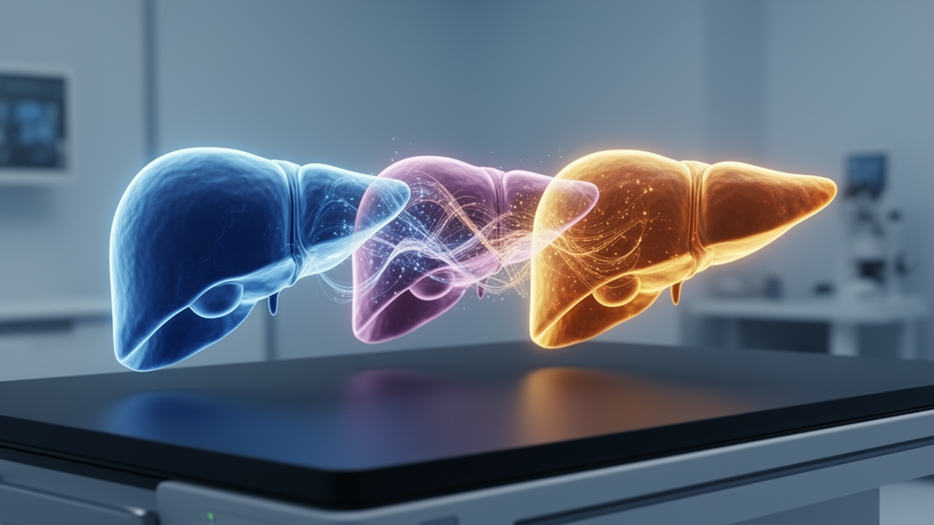
The technical rationale for avoiding co-registration is grounded in the physics and physiology of the acquisition. In gadoxetic acid-enhanced dynamic imaging, respiratory motion, variable breath-hold positions, and non-rigid liver deformation make reliable alignment across phases difficult, and any misalignment can propagate error directly into quantitative measurements. Instead of warping phases into alignment, the phase-adaptive model segments each phase independently, delineating liver parenchyma, intrahepatic non-parenchymal structures such as vessels, bile ducts, focal lesions, and post-treatment changes, and the spleen. Paraspinal muscle reference regions, needed for signal normalization, were obtained separately with TotalSegmentator. Parenchyma-only signal intensities from each phase then fed directly into calculations of arterial enhancement fraction, relative liver enhancement, hepatocellular uptake index, liver-to-spleen ratio, and liver-to-muscle ratio, with segmentation-derived organ volumes incorporated into index computation, particularly for the uptake index.
Performance on the independent test cohort of 500 annotated sequences from 100 patients was striking. Median Dice similarity coefficients for liver parenchyma ranged from 0.998 in the hepatobiliary phase to 0.999 in the other four phases, with median average surface distances below 0.04 millimeters. Spleen segmentation achieved near-perfect overlap, with a median Dice coefficient of 1.000 across all phases. Non-parenchymal liver structures, which are smaller and less conspicuously bounded, showed lower median Dice values of 0.873 to 0.945, peaking in the portal venous phase where contrast in the portal veins maximizes vessel-parenchyma differentiation. The phase-adaptive model outperformed TotalSegmentator MRI on both liver parenchyma and spleen across every phase. Mean inference time was 36 seconds per sequence and roughly three minutes per complete examination on a dedicated high-performance computing node.
Crucially, the automated indices agreed closely with what experienced human readers produce. In a radiological validation cohort of 50 patients, two radiology residents with five and four years of experience performed manual region-of-interest measurements using the local picture archiving system, placing standardized 20-square-millimeter regions on both liver lobes, the spleen, and the paraspinal muscles at the level of the spleen’s largest cross-section. Interreader agreement between the two radiologists was excellent for all five indices, with intraclass correlation coefficients of 0.960 to 0.989. Reader-model agreement was similar, ranging from 0.936 to 0.977, highest for the hepatocellular uptake index and lowest for the liver-to-muscle ratio. Bland-Altman analysis showed biases close to zero with narrow limits of agreement for relative liver enhancement, liver-to-spleen ratio, and liver-to-muscle ratio, while the uptake index and arterial enhancement fraction showed larger mean differences and wider limits, particularly in reader-model comparisons.
The clinical payoff emerged when the fully trained pipeline was applied to all 1,174 examinations in the clinical cohort. Patients categorized as cirrhotic — based on explicit documentation or radiology reports describing cirrhotic liver morphology — showed markedly lower hepatocyte-specific indices than non-cirrhotic patients: hepatocellular uptake index, liver-to-spleen ratio, normalized relative liver enhancement, and liver-to-muscle ratio were all significantly lower, with p-values below 0.001. The arterial enhancement fraction, which depends on early dynamic enhancement rather than hepatocyte-specific uptake, did not differ significantly between groups (p = 0.14). This pattern is consistent with reduced hepatocellular uptake of gadoxetic acid in chronic liver disease and demonstrates, at a scale that would be impractical with manual region-of-interest placement, that the automated indices behave as the underlying physiology predicts.
The authors are candid about what this approach does and does not improve. The primary advantage over conventional manual assessment lies in scalability, whole-liver parenchymal sampling, and reduced operator dependency rather than in any demonstrated index-level diagnostic superiority, which would require comparison against functional or physiological reference standards. Several limitations deserve emphasis. Training and testing occurred at a single center with consistent scanners and protocols, so multicenter external validation is warranted before generalizability can be assumed. Reference segmentations were generated by a semi-automatic annotation workflow followed by slice-by-slice manual correction, which may not provide the independence of fully de novo manual contouring — meaning the near-perfect Dice coefficients should be interpreted in light of that semi-automatic reference standard. No formal head-to-head diagnostic comparison between automated whole-liver and conventional region-of-based index calculation was performed, because a functional or histological reference standard was unavailable for the validation cohort.
Further caveats concern specific patient subgroups and reference signals. The muscle reference signal used for the liver-to-muscle ratio may be susceptible to pathological alterations such as sarcopenia, myopathy, or fatty infiltration, which were not systematically assessed. Indices requiring splenic normalization — the hepatocellular uptake index and liver-to-spleen ratio — could not be computed in patients with prior splenectomy, who were excluded from those analyses. And although the model was developed across a broad spectrum of hepatic pathologies, its performance may be limited where normal liver parenchyma is extensively replaced or anatomy is markedly distorted, as in diffuse infiltrative malignancy, polycystic liver disease, or after lobar resection; these scenarios were not systematically evaluated.
Even with those limitations, the study sketches a plausible route toward quantitative liver MRI at population scale. Because the pipeline runs on standard gadoxetic acid-enhanced acquisitions with no extra scanning, no co-registration, and no manual drawing, it could make multi-phase, multi-index liver quantification feasible in large clinical studies where manual measurement would be prohibitive. The authors note that further validation against clinical reference standards such as indocyanine green clearance, the LiMAx breath test, or composite scores like the Functional Liver Imaging Score — including head-to-head comparison of automated versus manual approaches — is needed to support clinical translation. For now, the message is a technical one: a single phase-adaptive model can segment the liver, its internal non-parenchymal structures, and the spleen across every phase of a dynamic contrast study, and the indices it computes track both expert readers and the expected biology of cirrhosis.
Subject of Research: Automated whole-liver quantification of signal intensity-derived liver indices from gadoxetic acid-enhanced MRI using phase-adaptive deep learning segmentation
Article Title: Phase-adaptive automated whole-liver quantification of signal intensity-derived liver indices from gadoxetic acid-enhanced MRI
Article References: Barajas Ordonez, F., Westfechtel, S., Matar, F., Schmidt, C., Lemessa, J., Wienholt, P., Truhn, D., Kuhl, C., Barabasch, A., & Nebelung, S. (2026). Phase-adaptive automated whole-liver quantification of signal intensity-derived liver indices from gadoxetic acid-enhanced MRI. European Radiology Experimental, 10(1), Article 155. https://doi.org/10.1186/s41747-026-00813-z
Image Credits: AI Generated
DOI: 10.1186/s41747-026-00813-z
Keywords: gadoxetic acid MRI, deep learning, liver segmentation, nnU-Net, cirrhosis, liver indices, hepatobiliary phase, radiology, quantitative imaging, medical imaging AI, Phase-adaptive, automated
News Source: Ophelia Keating. (October 29, 2026). AI Quantifies Liver Health Across All MRI Contrast Phases Without Registration. Scienmag.