In a groundbreaking advancement poised to transform lung cancer diagnostics, researchers have unveiled a cutting-edge machine learning model that predicts the invasiveness of lung adenocarcinoma manifesting as ground-glass nodules on CT scans. This innovation leverages the integration of sophisticated radiomics with clinical CT features, promising to elevate the precision of preoperative assessments and tailor therapeutic strategies with unprecedented accuracy.
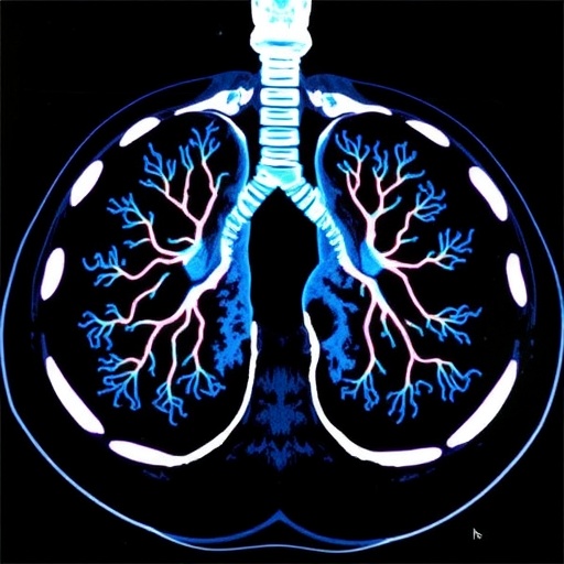
Lung adenocarcinoma, the predominant subtype of lung cancer, often presents diagnostically elusive characteristics on imaging—particularly when appearing as ground-glass nodules (GGNs). Conventional radiological methods, largely dependent on subjective interpretation, frequently struggle to differentiate between invasive and minimally invasive disease subtypes. This diagnostic uncertainty can significantly impact surgical planning and patient outcomes, underscoring the urgent need for more objective, robust tools in the clinical arsenal.
The research team embarked on a comprehensive, multicenter retrospective investigation involving 357 patients with pathologically confirmed lung adenocarcinoma. Their innovative approach combined high-resolution CT-derived radiomics and meticulously evaluated clinical imaging features, enabling the extraction of a vast repertoire of 1,129 radiomics parameters alongside 16 critical clinical CT attributes. These data-rich profiles formed the foundation for machine learning algorithms poised to redefine the boundaries of diagnostic capability.
Harnessing the power of principal component analysis (PCA) and the least absolute shrinkage and selection operator (LASSO) method for dimensionality reduction, the researchers distilled the immense feature set into the most salient predictors. This preprocessing step was crucial to mitigate overfitting and optimize model performance, effectively navigating the complexity of radiomic data to unveil the subtle imaging fingerprints of tumor behavior.
Five sophisticated machine learning classifiers were rigorously trained and evaluated: XGBoost, Support Vector Machine (SVM), Random Forest (RF), Logistic Regression, and Light Gradient Boosting Machine (LightGBM). Each model was fine-tuned to distinguish low invasiveness—comprising minimally invasive and Grade 1 invasive adenocarcinomas—from high invasiveness defined by Grades 2 and 3 invasive adenocarcinomas, thereby directly addressing the critical clinical stratification challenge.
Among these, the Random Forest model integrated with clinical CT features and PCA-transformed radiomics emerged as the superior predictive tool. Demonstrating an Area Under the Curve (AUC) of 0.854 on the training cohort, 0.769 on the test cohort, and maintaining robust performance with an AUC of 0.778 on an independent external validation set, the model’s consistency signals its potential for real-world clinical deployment.
Key predictive radiomic components elucidated by SHapley Additive exPlanations (SHAP) provided insightful interpretability to the model, empowering clinicians to understand the contributory impact of imaging features on invasion risk predictions. This transparency bridges the gap between complex algorithmic output and clinical decision-making, fostering trust and facilitating integration into routine practice.
This integrative model also significantly outperformed existing clinical-only models and a comparative clinical CT features-LASSO radiomics approach, illustrating the synergistic value of combining radiomic information with established clinical imaging data. Such enhanced predictive accuracy sets a new benchmark for non-invasive preoperative evaluation in lung cancer care.
The implications of this research extend beyond diagnostic accuracy. By augmenting early and precise identification of invasive lung adenocarcinoma, the model has the potential to guide nuanced surgical decisions, minimize unnecessary extensive resections, and personalize adjuvant therapy protocols—ultimately improving patient survival and quality of life.
While the study exemplifies a leap forward, the authors underscore the necessity of further validation through prospective, large-scale clinical trials. Such efforts would confirm the utility, generalizability, and cost-effectiveness of this technology across diverse populations and healthcare settings, ensuring the robustness of its clinical application.
Technically, this study embodies the confluence of radiomics—an emerging discipline that converts medical imaging into mineable high-dimensional data—and advanced machine learning algorithms capable of discerning complex patterns imperceptible to human observers. The methodology marks a paradigm shift, reinforcing the role of multidisciplinary innovation in tackling oncology’s diagnostic challenges.
Moreover, the reliance on dual-cohort validation, with an external dataset independent of training phases, strengthens the credibility of the findings. This design mitigates biases and ensures reproducibility—critical factors for transitioning such AI-driven models from research environments into clinical workflows.
The use of decision curve analysis further supplements the evaluation by assessing the clinical net benefit across varied threshold probabilities. This pragmatic metric accentuates the model’s potential clinical value beyond statistical indices, emphasizing its relevance for patient-centered care decisions.
In sum, the integration of radiomics with clinical CT features through robust machine learning not only enhances objectivity but also introduces a predictive precision previously unattainable through conventional radiological assessment alone. This nuanced approach fosters the evolution of personalized medicine paradigms in thoracic oncology.
As lung cancer remains a formidable global health burden, these advancements resonate profoundly with the ongoing quest to harness artificial intelligence for earlier, more precise detection and treatment stratification. The research anchors future opportunities for interdisciplinary collaboration at the intersection of radiology, oncology, and computational science.
The full promise of this technology lies in its scalability and adaptability to varied imaging platforms and patient demographics, which future research must rigorously explore. Nevertheless, this study lays a strong foundation indicating that AI-empowered radiomics can indeed advance the frontier of lung cancer diagnostics.
Ultimately, embracing such innovative diagnostic tools heralds a transformative phase in cancer care, where data-driven insights augment clinical expertise to deliver personalized, effective interventions with greater confidence and improved patient outcomes.
Subject of Research:
Machine learning-based prediction of invasiveness in lung adenocarcinoma presenting as ground-glass nodules using radiomics and clinical CT features.
Article Title:
Machine learning-based prediction of invasiveness in lung adenocarcinoma presenting as ground-glass nodules using radiomics and clinical CT features
Article References:
Lin, M., Li, L., Hui, Y. et al. Machine learning-based prediction of invasiveness in lung adenocarcinoma presenting as ground-glass nodules using radiomics and clinical CT features. BMC Cancer 25, 1693 (2025). https://doi.org/10.1186/s12885-025-14983-3
Image Credits:
Scienmag.com
DOI:
03 November 2025
Tags: advanced CT imaging techniquesAI in lung cancer diagnosticsclinical imaging features in oncologydiagnostic uncertainty in lung cancerground-glass nodules on CT scanslung adenocarcinoma invasiveness detectionmachine learning for cancer predictionmulticenter study on lung cancerobjective tools for cancer diagnosispatient outcomes in lung adenocarcinomapreoperative assessment in lung cancerradiomics in medical imaging


