In a groundbreaking advance poised to reshape our understanding of cancer genesis, researchers at EMBL Heidelberg have unveiled an innovative AI-powered technology that deciphers the elusive origins of chromosomal instability. This instability, a hallmark of many aggressive cancers, involves numerical and structural abnormalities in chromosomes that compromise genetic integrity and spearhead malignant transformation. The new tool, dubbed MAGIC—short for machine learning-assisted genomics and imaging convergence—heralds a new era in cellular analysis by seamlessly integrating automated microscopy, advanced AI algorithms, and genomic sequencing. Through this fusion, MAGIC can unerringly detect and label rare cellular anomalies with unprecedented precision and scale, offering insights that were previously unattainable with conventional methods.
Cancer has long been recognized as a disease rooted in genetic chaos. Mutations, chromosomal breaks, and irregular rearrangements precipitate a breakdown in normal cellular function, leading cells to evade growth controls and proliferate uncontrollably. For well over a century, scientists have hypothesized the critical role of chromosomal abnormalities in this process, dating back to Theodor Boveri’s early microscopy observations in the early 1900s. Yet, capturing these chromosomal aberrations in living cell populations has remained a formidable challenge. Cells harboring such defects are typically scant in number and prone to elimination through natural selection mechanisms, rendering their identification akin to finding needles in a cellular haystack.
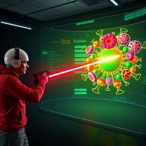
MAGIC revolutionizes this pursuit by automating what was previously a labor-intensive and error-prone task. It deploys a sophisticated machine learning model trained on manually annotated images to recognize a telltale cellular feature known as the micronucleus. These diminutive, DNA-containing compartments detach from the main nucleus and are a definitive sign of underlying chromosomal instability. By effectively performing a digital version of laser tag, MAGIC directs a laser beam to “tag” these micronucleated cells through a photoconvertible dye. This dye alters its fluorescence upon exposure to targeted light, enabling precise marking of those cells for subsequent isolation and study without disrupting their viability.
The ramifications of this technology are profound. Through high-throughput automated microscopy paired with AI-driven image analysis, MAGIC can analyze tens of thousands of cells within a single day, a feat unattainable by manual microscopy. This scale facilitates robust statistical assessments of the frequency and causes of chromosomal abnormalities, providing a window into the cell division dynamics that foster genomic instability. Early usage of MAGIC has revealed that over 10% of cell divisions result in spontaneous chromosomal errors. Strikingly, this incidence nearly doubles in cells where the tumor suppressor gene p53 is mutated—a frequent mutation in human cancers—highlighting the gene’s pivotal role in maintaining chromosomal fidelity.
Furthermore, MAGIC’s insights extend beyond mere rate quantification. By correlating micronucleus presence with locations of DNA double-strand breaks, it sheds light on the genomic landscapes susceptible to instability. This coupling of imaging and genomics offers a multidimensional perspective crucial for unraveling the mechanistic underpinnings that drive chromosomal missegregation and rearrangement during mitosis. These insights could illuminate pathways leading to metastasis, drug resistance, and tumor relapse, which are tightly linked to chromosomal instability.
The development of MAGIC epitomizes interdisciplinary collaboration, uniting expertise across computer vision, robotic automation, genomics, and microscopy. The core team from EMBL Heidelberg worked closely with the Advanced Light Microscopy Facility and partners at the German Cancer Research Centre, among others, to engineer this powerful platform. Their efforts exemplify how melding cutting-edge AI with biological research can surmount previously insurmountable obstacles in cell biology.
Looking ahead, the flexibility of MAGIC promises broad applicability. Although trained to detect micronuclei in this inaugural study, the underlying machine learning algorithms can theoretically be adapted to identify diverse cellular features indicative of pathogenic or physiological states. Hence, this platform could become an indispensable tool across numerous biological disciplines, from neuroscience to immunology, wherever visual cell phenotyping and subsequent molecular characterization are needed.
Importantly, MAGIC is not merely a tool for cancer researchers; it represents a paradigm shift in how we perform single-cell analysis. By automating detection and marking at a cellular level in living populations, it bridges the gap between high-content imaging and genomic interrogation, enabling unprecedented resolution in studying cellular heterogeneity. This technological leap will expedite discovery and, ultimately, foster new diagnostic and therapeutic strategies targeting chromosomal instability’s root causes.
In essence, MAGIC embodies the convergence of artificial intelligence and biological microscopy, overcoming longstanding technical hurdles in cancer biology. By illuminating the origins and dynamics of chromosomal instability with unprecedented clarity and throughput, it provides a vital new lens through which to explore cancer’s earliest and most consequential genetic derangements. As malignancies continue to challenge human health globally, such innovations hold promise not only to deepen scientific understanding but also to spur development of interventions that intercept cancer at its genomic inception.
European Molecular Biology Laboratory’s senior scientist Jan Korbel, who led the study published in Nature, emphasizes that this technology aligns with the cutting edge of AI-driven biology: “Our system can be trained on almost any visually distinguishable cellular trait, opening new vistas for biological exploration and discovery.” This union of machine learning, robotics, and genomics stands as a testament to the transformative potential of interdisciplinary science in tackling humanity’s most daunting diseases.
Subject of Research: Cells
Article Title: Origins of chromosome instability unveiled by coupled imaging and genomics
News Publication Date: 29-Oct-2025
Web References: DOI: 10.1038/s41586-025-09632-5
Image Credits: Daniela Velasco/EMBL
Keywords: Molecular biology
Tags: advanced cellular analysis technologyAI-powered cancer researchautomated microscopy in cancer studiescancer origins and mutationschromosomal instability detectionEMBL Heidelberg research innovationsgenomic sequencing advancementsinnovative cancer diagnosis toolsmachine learning in genomicsprecision medicine in oncologyrare cellular anomaly identificationTheodor Boveri cancer theories
