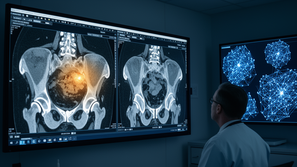
Ovarian cancer remains one of the most elusive killers in oncology. It is the eighth most common cancer in women and the deadliest gynecological malignancy, yet it is rarely caught early because its symptoms are vague or absent until the disease has advanced. Now, a team of researchers in Romania has developed an artificial intelligence system that reads CT scans with remarkable precision, tracing the outlines of ovarian tumors pixel by pixel and sorting them into molecularly defined categories — a technical feat that could one day help radiologists and oncologists make faster, better-informed decisions.
The study, published in Frontiers in Artificial Intelligence by Miruna-Cristina Andone, Loretta Ichim, and Dan Popescu of the National University of Science and Technology Politehnica of Bucharest, introduces a two-stage neural network fusion system. Rather than relying on a single deep learning model, the approach combines the strengths of several architectures in a layered ensemble, achieving an average F1 score of 93.53 percent across five classes, with recall of 93.38 percent and precision of 93.68 percent on an independent test cohort. Those numbers outperformed every individual network and every first-stage ensemble tested in the study.
The stakes are enormous. According to data cited in the paper, roughly 324,603 women were diagnosed with ovarian cancer in 2022, and the World Ovarian Cancer Coalition projects that four million lives will be lost to the disease by 2040, with approximately 12 million new cases diagnosed by 2050. Epithelial ovarian cancer accounts for about 95 percent of ovarian malignancies, and the five-year survival rate for late-stage diagnoses remains below 30 percent. Because the disease is often detected only after it has metastasized, when surgery may no longer be possible, earlier and more accurate imaging-based detection is a global health priority.
Traditional diagnosis and staging rely on manual analysis of CT and MRI scans, a process that is subjective, expensive, and prone to human error. Deep learning has begun to change that calculus. Neural networks can highlight tumor structures of various sizes, including small lesions that might escape a tired eye, and radiomic analysis can extract quantitative features that extend beyond what a radiologist sees. But individual models have weaknesses — they can be brittle, sensitive to noise, and inconsistent across varied data. Ensembles, which combine multiple models, offer a way to compensate for those deficiencies.
The Romanian team tested nine well-known architectures on the TCGA-OV dataset, a public collection from The Cancer Imaging Archive comprising 143 patients and 26,570 CT images. The architectures included U-Net, Attention U-Net, SegNet, ResNet50, VGG16, GoogLeNet, DenseNet-121, DeepLabV3+, and the Vision Transformer (ViT). Each was adapted for pixel-wise semantic segmentation, with classification-oriented networks stripped of their classification heads and fitted with segmentation decoders. The researchers then ranked the models using F1 score, recall, and precision for each of five classes, and selected the top three performers: Attention U-Net, DeepLabV3+, and ViT.
Those three networks bring complementary skills. Attention U-Net, a variant of the classic U-shaped encoder-decoder, uses attention mechanisms to focus on relevant regions of the image, sharpening its sensitivity to tumor edges and abnormal structures. DeepLabV3+ combines atrous — or dilated — convolutions with an effective decoder to capture contextual information at multiple spatial scales, excelling at boundary delineation. ViT, the newcomer among them, treats images as sequences of patches and uses self-attention to learn global relationships between distant regions, a capability that convolutional networks lack. Together, they cover local detail, contextual scale, and long-range coherence.
The first stage of fusion builds four ensembles from these three networks. Ensemble E1 uses binary voting, where each network casts a simple yes-or-no decision for each class. Ensembles E2, E3, and E4 use weighted scores based on F1, recall, and precision respectively, with weights assigned per network and per class. The second stage then fuses the outputs of all four ensembles into a Global System, weighted by the F1 scores of the first-level ensembles. At every step, the final classification of a pixel is determined by the highest weighted score across the five classes: No Tumor, Differentiated, Proliferative, Mesenchymal, and Immunoreactive.
One important nuance deserves attention. The four tumor classes beyond No Tumor are not read directly from the CT image. Instead, they are patient-level molecular subtype labels from The Cancer Genome Atlas genomic classifications, mapped onto the anatomically annotated tumor masks. The authors are explicit that this five-class formulation should not be interpreted as voxel-level prediction of molecular subtypes from imaging alone. Rather, the framework evaluates how accurately the models reproduce these mapped labels within segmented tumor regions, combining anatomical segmentation with subtype-derived labeling for multi-class analysis.
The experimental results show a clear progression. The three selected individual networks achieved average F1 scores between 86.01 and 86.59 percent on the test set. The first-level ensembles pushed that range to between 90.40 and 90.50 percent. The Global System then reached 93.53 percent. Patient-level analysis, which treats each subject as a single unit to account for the correlation among slices from the same patient, confirmed the pattern: the Global System achieved a mean patient-level F1 score of 93.5 ± 2.4 percent, compared with 91.8 percent for Attention U-Net, 91.5 percent for DeepLabV3+, and 92.1 percent for ViT. Paired Wilcoxon signed-rank tests showed the differences were statistically significant, with p-values ranging from 0.008 to 0.021.
The authors are careful about limitations. The testing cohort contains a limited number of independent subjects, and the same validation cohort was used both to select the component networks and to derive ensemble weights, which may introduce optimization bias. The study is a retrospective methodological evaluation on a public dataset, not a clinical validation. Boundary-based metrics such as Hausdorff distance were outside the predefined protocol. Still, the two-stage fusion framework demonstrates technical feasibility and points toward a future where multimodal integration — combining imaging with histopathology and genomic data — could sharpen diagnostic accuracy further. For a disease that claims roughly 150,000 lives annually, every percentage point of precision matters.
Subject of Research: Two-stage neural network fusion for ovarian cancer tumor segmentation from CT images
Article Title: Segmentation of ovarian cancer tumors from CT images based on a two-stage neural network fusion system
Article References: Andone, M.-C., Ichim, L., & Popescu, D. (2026). Segmentation of ovarian cancer tumors from CT images based on a two-stage neural network fusion system. Frontiers in Artificial Intelligence, 9, Article 1974931. https://doi.org/10.3389/frai.2026.1974931
Image Credits: AI Generated
DOI: 10.3389/frai.2026.1974931
Keywords: ovarian cancer, artificial intelligence, medical imaging, CT segmentation, neural networks, deep learning, ensemble models, TCGA-OV, Segmentation, ovarian, cancer, tumors
News Source: Nathaniel Bowman. (October 16, 2026). AI Fusion System Sharpens Ovarian Tumor Segmentation From CT Scans. Scienmag.