A groundbreaking advancement in the diagnosis of coal workers’ pneumoconiosis (CWP), a chronic occupational lung disease notorious for its complex and irreversible pulmonary complications, has emerged from the integration of deep learning with high-resolution computed tomography (HRCT) imaging. Researchers have developed a sophisticated algorithm that offers unparalleled accuracy in classifying clinical imaging features, potentially revolutionizing how this debilitating disease is identified and managed in clinical settings.
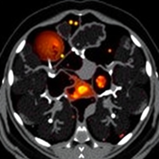
Coal workers’ pneumoconiosis, often known as “black lung disease,” represents a severe health hazard for miners exposed to coal dust. Traditional diagnostic methods have heavily relied on chest X-rays, which struggle to capture the nuanced and intricate lung changes associated with CWP. This limitation has hindered timely and accurate diagnosis. To overcome this, the new research pivots towards analyzing high-resolution computed tomography images, which provide more detailed views of pulmonary structures, ensuring subtle pathological changes do not go unnoticed.
The central innovation lies in leveraging a cutting-edge deep learning model called DenseNet combined with an Efficient Channel Attention Network (ECA-Net). This hybrid model exploits the intricate patterns and spatial hierarchies in HRCT images, enabling the effective distinction between different clinical manifestations of pneumoconiosis. The model was trained using an extensive dataset gathered from 217 patients with confirmed CWP and dust-exposed workers, allowing it to learn and recognize the complex imaging signatures unique to this disease.
The research team painstakingly annotated more than 1700 regions of interest (ROIs) within the HRCT images, categorizing them into four distinct clinical imaging features. These categories include small miliary opacities, nodular opacities, interstitial changes, and emphysema, each representing different pathological patterns indicative of disease severity and progression. By incorporating a robust data augmentation strategy, the researchers enhanced the dataset’s diversity, enabling the model to generalize well and maintain high performance across varying imaging scenarios.
In rigorous testing utilizing tenfold cross-validation, the DenseNet-ECA model achieved an extraordinarily high average area under the receiver operating characteristic curve (AUC) of 0.98, demonstrating exceptional discriminatory power. Remarkably, each imaging feature was classified with an AUC exceeding 0.92, underscoring the model’s consistent precision. Nodular opacities and emphysema, in particular, were identified flawlessly with AUCs of 1.0, reflecting zero classification errors in these categories.
Beyond technical prowess, the practical implications for clinical radiology are profound. Automated, reliable classification of HRCT images can provide radiologists with invaluable diagnostic support, reducing human error and diagnostic time. This is particularly critical in regions burdened with occupational lung diseases where specialist expertise may be limited. The algorithm’s ability to discern subtle imaging variations promises to improve early detection, monitor disease progression, and tailor intervention strategies more effectively.
Furthermore, the use of HRCT imaging mitigates the diagnostic ambiguity often encountered in chest X-rays, where overlapping anatomical structures obscure lung details. However, the sheer volume and complexity of HRCT images typically demand considerable time and expertise for manual interpretation. This novel algorithm addresses this bottleneck by automating classification, enhancing diagnostic throughput, and allowing clinicians to focus on patient care and treatment optimization.
While the study primarily focuses on CWP, the framework’s flexibility suggests broader applications across other interstitial lung diseases and occupational respiratory conditions. The research opens avenues for exploring similar deep learning approaches in diseases where imaging plays a pivotal role but remains challenging due to complex presentation patterns. This could spearhead a new era of AI-assisted diagnostics in pulmonary medicine.
Importantly, the researchers underline that their approach does not replace clinical judgment but rather augments it. By providing highly accurate, transparent, and reproducible assessments of HRCT features, the tool acts as a second set of eyes, enabling radiologists to confirm their impressions or identify findings that might otherwise be overlooked. This synergy between AI and human expertise exemplifies the future direction of medical imaging diagnostics.
The successful trial of this DenseNet-ECA model was registered with the Chinese Clinical Trial Registry, underscoring its methodological rigor and clinical relevance. The registration details further enhance transparency and encourage future research collaborations aimed at refining and validating the model across diverse populations and imaging modalities.
As occupational health continues to grapple with the consequences of industrial exposure, innovations such as this deep learning-based classification algorithm herald transformative progress. By harnessing artificial intelligence and high-resolution imaging, the medical community moves closer to eradicating diagnostic uncertainty, ultimately improving outcomes for countless individuals affected by coal workers’ pneumoconiosis worldwide.
This study, published in the prestigious journal BioMedical Engineering OnLine, not only exemplifies interdisciplinary collaboration between engineering and medicine but also spotlights the pivotal role of AI in addressing complex healthcare challenges. With continued development and integration into clinical workflows, such technologies promise more personalized, timely, and accurate diagnostic processes, reshaping the future landscape of pulmonary disease management.
Subject of Research: Deep learning-based classification of high-resolution computed tomography features in coal workers’ pneumoconiosis
Article Title: Deep learning-based algorithm for classifying high-resolution computed tomography features in coal workers’ pneumoconiosis
Article References:
Dong, H., Zhu, B., Kong, X. et al. Deep learning-based algorithm for classifying high-resolution computed tomography features in coal workers’ pneumoconiosis. BioMed Eng OnLine 24, 7 (2025). https://doi.org/10.1186/s12938-025-01333-4
Image Credits: Scienmag.com
DOI: https://doi.org/10.1186/s12938-025-01333-4
Tags: advanced diagnostic algorithmsAI in medical imagingblack lung disease identificationchronic occupational lung diseasecoal workers’ pneumoconiosis diagnosisdeep learning in healthcareDenseNet deep learning modelEfficient Channel Attention Networkhealthcare technology innovationshigh-resolution CT scansimproving diagnostic accuracy in CWPpulmonary disease classification


