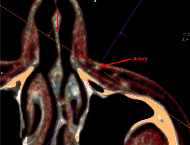
Credit: Victor Gombolevskiy et al. / Aesthetic Surgery Journal 2020
Researchers from the Center for Diagnostics and Telemedicine together with colleagues from Mayo Clinic College of Medicine and Science, University of Munich and Sechenov University used computed tomography to analyze the individual anatomy of the nasolabial triangle. They identified possible options for the distribution of blood vessels on three-dimensional course. It will help in safety planning of plastic and reconstructive surgeries and procedures. Study results published recently in the Aesthetic Surgery Journal, added to the series of publications about the aesthetic medicine project, which was launched by the Center for Diagnostics and Telemedicine in collaboration with Mayo Clinic.
The anatomy of each person is unique. It is important to know its details in order not to harm a patient during any medical procedure. Special attention always should be paid to the nasolabial triangle.
This part of the face is bounded by the nose on the top, by lips from below, and on the sides by nasolabial folds. In aesthetic medicine, it is one of the most challenging facial regions, since there is a dense network of blood vessels, both arterial and venous. Since with aging, nasolabial sulcus become more pronounced, cosmetic procedures are often performed on this area, such as injections of hyaluronic acid, which fills the subcutaneous space, and evens out the fold. However, ignorance of details of the individual vascular anatomy of this area can result in serious complications, including loss of vision.
In the depth of the nasolabial sulcus, the angular artery is located which is the terminal branch of the facial artery, which supplies blood to the most part of the face. Although its anatomy is well studied, there is a lack of information on how deep the angular artery is located relatively to the skin surface, as well as how its location can vary in three dimensions depending on age, gender and body mass index (BMI). The researchers decided to fill in this gap for people of the Caucasian ethnic group.
“We hope that clearer understanding of the pathway of the artery in this high-risk area will help make surgical procedures safer,” said the author Victor Gombolevsky, Head of the Department of Radiology Quality Development at the Center for Diagnostics and Telemedicine.
The research was based on analyses of cranial CT scans from the radiology database of the Center for Diagnostics and Telemedicine. Researchers analyzed CT scans of 150 patients of different age – from 14 to 89 years old, and with various BMI – from 16.7 to 47.8 kg/m2. Their nasolabial sulcus were examined on both sides in three dimensions.
In the entire sample, researchers described three types for angular arteries. In most cases (90%), one arterial trunk was found, the trunk bifurcated in 9%, and three trunks were discovered in 1% of the investigated cases. On average, the angular artery was located at the depth of 21.6 mm in the nasolabial fold, and at a depth of 8.9 mm in the nasal area. In 100% of cases, the angular artery located laterally to the nasolabial sulcus (i.e. closer to the cheek).
It is interesting that in contrast to the prevailing data, we discovered that the depth of the artery varied, and it was located most superficially in the area of the lip junction. In addition, in all cases the artery was located to the side of the nasolabial sulcus at variable distance from it with a greater difference in the nasal area. It turned out that gender and BMI did not have a significant impact on these facts, however, it was a considerable influence of age.
“With aging, both the depth of the angular artery and lateral distances between the nasolabial sulcus and the artery decrease significantly. This emphasizes the need for special caution when injecting soft tissue fillers, such as hyaluronic acid, into the furrow to elderly patients. Loss of fat volume of facial tissues exposes arteries, and it is much easier to damage them during injection procedures, “the authors say.
###
Media Contact
Stanislav Samburskiy
[email protected]
Original Source
https:/
Related Journal Article
http://dx.


