Artificial intelligence has transformed the interpretation of adult medical images, yet its reach into the smallest patients has been strikingly limited. Fewer than one in five commercially available AI models are cleared for paediatric use, and only about 17 per cent of commercial models have received approval from the US Food and Drug Administration for paediatric patients. The reasons are structural rather than technical: children make up less than one per cent of public medical imaging datasets, privacy regulations constrain data sharing between hospitals, and curating and labelling paediatric images demands clinical effort that most single institutions cannot spare. The result is a field in which most paediatric AI models are built on single-centre data, few report external validation, and many lose accuracy the moment they cross an institutional boundary. A new international study, published in Nature Biomedical Engineering, shows that a deceptively simple training strategy can break this deadlock.
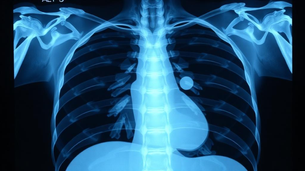
The research, led by Vishwanatha M. Rao, Emma Chen and Agustina Saenz at Harvard Medical School under the supervision of Pranav Rajpurkar, assembled 30 neonatal intensive care units across 20 countries and territories into one of the most geographically diverse imaging cohorts ever gathered for neonatal AI development. Their target task was the assessment of endotracheal tubes on chest radiographs, a deliberately chosen proof of concept. Several AI tools have already received FDA clearance for checking tube position in adult intensive care, but no commercial solution exists for newborns, despite the fact that misplaced tubes in neonates carry severe consequences: tubes inserted too deeply can ventilate a single lung and cause collapse, while false alarms can trigger unnecessary re-intubations that raise procedural risk and haemodynamic instability.
The team’s method is called continual learning, and it works like a relay race for model weights. A deep learning model originally trained on adult ICU radiographs was passed sequentially from hospital to hospital. Each site retrained the model on just 50 locally curated neonatal chest X-rays inside its own computing environment, then forwarded only the updated parameters to the next institution. No patient image ever left a hospital, no large dataset had to be assembled, and no specialized AI team was required at any site. Because only model weights travel, the approach sidesteps the data transfer agreements, ethics approvals across jurisdictions and regulatory hurdles that make centralized data pooling so difficult, while avoiding the synchronized compute infrastructure and multi-round communication that federated learning demands.
Technically, the underlying model was CarinaNet, a RetinaNet-based object detector with a ResNet-50 backbone and a feature pyramid network, pretrained on natural images and then adapted to 1,890 adult ICU radiographs. The model detects two structures, the tip of the endotracheal tube and the carina, the cartilaginous ridge where the trachea splits into the bronchi, and outputs bounding boxes whose centre points serve as predicted landmark locations. Its training loss combines focal loss for classification with smooth L1 regression, weighted asymmetrically to prioritize the carina, the anatomical reference point for judging tube depth. Images were histogram-equalized to standardize contrast across scanners, resized to 640 by 640 pixels with aspect-ratio-preserving padding, and augmented with small random rotations and vertical crops.
The results were decisive. For endotracheal tube tip localization, median error fell from 2.35 centimetres when the adult model was applied to neonatal images without adaptation, to 0.94 centimetres with hospital-specific fine-tuning, and down to 0.40 centimetres with continual learning. Carina localization improved from 1.62 centimetres under direct prediction and 0.27 centimetres under fine-tuning to 0.20 centimetres. Combined, the continual learning models achieved a mean landmark error of 0.67 centimetres, compared with 1.29 centimetres for fine-tuning and 4.30 centimetres for the unadapted adult model, improving localization at 29 of 30 sites, with statistical significance at 27. The study cohort comprised 2,965 radiographs from 2,960 newborns, with a median age at imaging of just two days, a mean gestational age of 32.9 weeks and a mean birth weight of 2,195 grams, reflecting a predominantly preterm population.
The clinical stakes of those millimetres are considerable. The researchers defined extreme errors as combined localization mistakes exceeding five centimetres, a threshold informed by anatomical measurements showing that the total tracheal length in neonates ranges from roughly 2.2 to 6.4 centimetres depending on gestational age. A five-centimetre error would therefore approximate or exceed the entire trachea of most newborns. Continual learning cut the rate of such catastrophic errors to 2.6 per cent of cases on average, against 8.2 per cent for fine-tuning and a startling 37.4 per cent for direct prediction. When the models were asked to triage, ranking the fifteen most concerning scans per hospital for radiologist review, continual learning achieved significantly higher precision and recall for detecting abnormally low tube placements than fine-tuning at 18 of 30 hospitals, with positive improvements at 27, and precision exceeding 80 per cent at the best-performing sites.
A crucial question for any sequential training scheme is catastrophic forgetting, the tendency of a model to lose earlier skills as it absorbs new data. The team addressed this with a backward evaluation: across 100 random hospital orderings, they compared each hospital’s performance immediately after the model trained on its data with performance after the full sequence completed. Far from degrading, the final model consistently outperformed the intermediate one at hospitals encountered early in the sequence, with a mean improvement of 0.48 centimetres in combined error at the first position. Knowledge, in other words, accumulated rather than eroded, and the gains grew steadily with each additional contributing hospital across the entire 29-hospital training sequence, with no evidence of a plateau.
The study also mapped the conditions under which the paradigm succeeds. Initializing the sequence with the adult-pretrained model beat starting from a generic object detector never exposed to radiographs, improving localization at 25 of 30 sites and triage metrics at 27, confirming that features learned from adult chests transfer usefully to newborns despite profound anatomical differences. But data quality mattered sharply: one contributing hospital supplied notably low-resolution radiographs, and including those scans in training raised the combined error from 0.67 to 0.75 centimetres and reduced the number of sites with positive triage improvements from 27 to 20, suggesting that quality thresholds for participating sites may be warranted. Encouragingly, performance remained consistent across demographic subgroups of race, sex, age, birth weight and gestational age, with no significant pairwise differences.
The researchers are candid about the limits. Pixel-to-centimetre conversions relied on the known width of the endotracheal tube as an internal calibration reference, an imperfect proxy that probably explains weaker correlations between predicted distances and clinical labels for high tube placements, which are properly defined relative to the first thoracic vertebra rather than the carina. The retrospective design simulated decentralized training centrally, and true distributed execution, prospective trials measuring time-to-confirmation and re-intubation rates, and reconciliation with regulatory oversight of continuously evolving models all remain ahead. Yet the broader implication is hard to overstate. Any paediatric imaging task for which adult AI models exist but paediatric solutions do not could, in principle, be reached the same way: a modest locally annotated dataset at each hospital, a self-contained training container, and a travelling model that grows wiser with every stop. In a field where children have been systematically left out of the AI revolution, thirty hospitals across twenty countries have just shown how to bring them in, one small, private dataset at a time.
Subject of Research: Privacy-preserving continual learning to adapt adult AI radiograph models for neonatal endotracheal tube assessment
Article Title: Repurposing adult AI radiograph models for neonatal care through continual learning: an international multicentre study
Article References: Repurposing adult AI radiograph models for neonatal care through continual learning: an international multicentre study. (n.d.). https://doi.org/10.1038/s41551-026-01812-6
Image Credits: AI Generated
DOI: 10.1038/s41551-026-01812-6
Keywords: continual learning, neonatal intensive care, chest radiographs, endotracheal tube placement, medical AI, privacy-preserving machine learning, catastrophic forgetting, paediatric radiology, multicentre study, transfer learning, deep learning, Nature Biomedical Engineering
News Source: Harold Sullivan. (October 13, 2026). Adult AI Learns to Read Newborn Chest X-rays by Travelling Between 30 Hospitals. Scienmag.