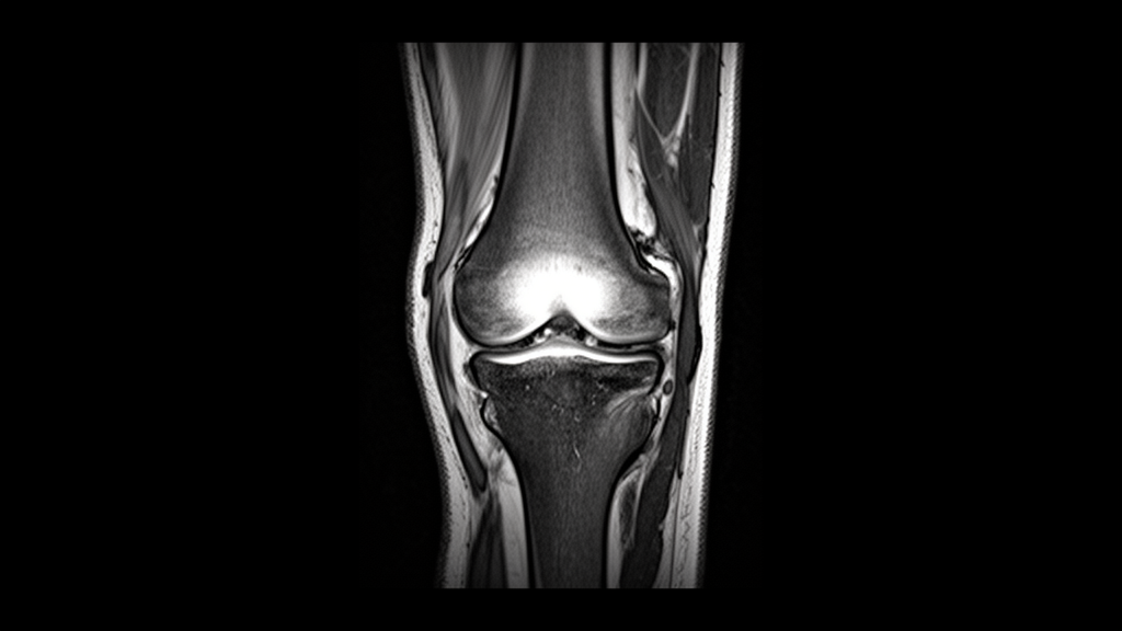
At Aarupadai Veedu Medical College and Hospital in Puducherry, an intra-individual comparative imaging study evaluating seventy-two patients undergoing knee magnetic resonance imaging established that spectral adiabatic inversion recovery consistently produced more homogeneous fat suppression and superior anatomical visualization compared with spectral pre-saturation with inversion recovery. Conducted from March to August 2025 on a 1.5-Tesla Philips Achieva scanner, the paired trial examined whether adiabatic radiofrequency pulses could overcome the field inhomogeneity challenges commonly observed in peripheral musculoskeletal imaging. Across seven distinct joint structures—including the cruciate ligaments, meniscal fibrocartilage, articular cartilage, patella, tendons, femoral condyles, and Hoffa’s fat pad—proton density-weighted sequences utilizing the adiabatic technique demonstrated statistically significant improvements in qualitative clarity over conventional pre-saturation sequences. Two blinded musculoskeletal radiologists independently assessed the paired imaging datasets, recording significant enhancements under both nonparametric Wilcoxon signed-rank tests and secondary parametric paired t-tests, with probability values falling below one-thousandth across every evaluated anatomical region.
Magnetic resonance imaging represents the noninvasive gold standard for evaluating internal derangements of the human knee joint, maintaining high-contrast resolution and multiplanar capabilities without exposing patients to ionizing radiation since its initial knee applications in 1983. The knee joint functions as a mechanically intricate articulation stabilized by the anterior cruciate ligament, posterior cruciate ligament, medial collateral ligament, lateral collateral ligament, and patellar ligament, all of which are situated in close physical proximity to dense adipose tissue deposits. When routine imaging sequences are acquired without fat suppression, the intense signal emitted by intra-articular and periarticular fat—most notably within Hoffa’s fat pad—frequently masks subtle ligamentous tears, meniscal disruptions, and chondral flaws. Furthermore, accompanying pathological alterations such as joint effusion substantially modify intra-articular signal characteristics, reinforcing the requirement for precise, uniform fat suppression to allow dependable diagnostic differentiation between normal anatomical boundaries and pathological derangements during routine clinical evaluation.
Spectral pre-saturation with inversion recovery operates as a chemical-shift-selective approach originally designed for spinal examinations before gaining broad acceptance across musculoskeletal imaging protocols. The technique utilizes a frequency-specific one-hundred-and-eighty-degree radiofrequency inversion pulse calibrated specifically to the resonance frequency of fat protons, which are shifted by approximately 224 Hertz relative to water protons at a 1.5-Tesla field strength. After applying this radiofrequency pulse, longitudinal magnetization flips, and image acquisition is timed to initiate precisely when fat proton magnetization passes through its null point along the longitudinal axis, thereby silencing the lipid signal while preserving water-containing tissue signal. In contrast to broad short tau inversion recovery methods, this selective approach leaves non-lipid short longitudinal relaxation tissues unaffected, preserving both post-contrast enhancement and higher signal-to-noise ratios. However, the sequence remains inherently vulnerable to main magnetic field and radiofrequency field inhomogeneities, which precipitate frequency misregistration, incomplete suppression, and localized artifacts near curved peripheral joint contours.
Spectral adiabatic inversion recovery functions as a sophisticated hybrid technique that integrates the chemical shift selectivity of saturation methods with the inversion stability of adiabatic radiofrequency pulses. Because the adiabatic pulse is frequency-modulated to invert solely fat protons, the sequence selectively extinguishes the lipid component while protecting water tissue signal and maintaining adequate signal-to-noise ratios throughout the imaging volume. In addition, the method employs non-slice-selective inversion pulses, which notably reduces inter-slice cross-talk and minimizes artifacts caused by patient motion across multi-slice acquisitions. Although adiabatic pulses inevitably generate a higher specific absorption rate and demand slightly longer acquisition intervals, their inherent resistance to radiofrequency field variations provides an optimized theoretical foundation for imaging complex, curved anatomical regions such as the human knee.
Although the physical principles explaining the resistance of adiabatic pulses to magnetic field and radiofrequency inhomogeneities have been established, direct intra-individual comparisons detailing structure-specific clarity within knee imaging had remained sparse, with previous comparative evaluations focusing primarily on the wrist, spine, or sacroiliac joint. To characterize these comparative differences in the knee, the investigators designed a prospective, intra-individual investigation involving seventy-two consecutive patients referred for diagnostic knee examinations at their tertiary care academic hospital in South India. Purposive sampling was used to enroll participants presenting with typical clinical indications, including joint pain, swelling, suspected ligamentous or meniscal disruptions, and articular cartilage pathology. The research protocol, consent forms, and data collection procedures received formal ethical clearance from the Institutional Human Ethics Committee, and all adult subjects provided written informed consent, with parental consent and patient assent obtained for individuals under eighteen years of age.
The recruited patient cohort encompassed forty-seven male participants, representing 65.3 percent of the study group, and twenty-five female participants, representing 34.7 percent, with ages ranging from ten to sixty-nine years and the highest concentration situated within the twenty to thirty-nine age bracket. Candidate participants were systematically excluded if they possessed metallic implants or orthopedic prostheses near the knee, suffered from claustrophobia, had a history of previous knee surgery, exhibited severe motion artifacts that rendered scans non-diagnostic, or failed to complete sequences due to technical interruption. Each patient underwent examination on the identical 1.5-Tesla scanner utilizing a dedicated knee coil, lying supine with the mid-knee joint centered at the magnetic isocenter. The imaging protocol paired routine sagittal, coronal, and axial proton density-weighted pre-saturation sequences and turbo spin-echo sequences with an experimental sagittal adiabatic sequence acquired during the same examination session, strictly standardizing repetition time, echo time, field of view, slice thickness, matrix size, bandwidth, and number of excitations.
To eliminate potential evaluation bias, all acquired image series were anonymized and transferred to picture archiving and communication systems in randomized order before assessment by two independent musculoskeletal radiologists. The primary evaluator, an associate professor possessing over eight years of specialized radiology experience, and the secondary evaluator, an assistant professor with more than three years of experience, performed their reviews blinded to patient identities, sequence allocations, and each other’s scores. Both radiologists independently graded fat suppression homogeneity, the presence of technical artifacts, and anatomical visualization across seven predetermined joint structures: the femoral condyles, surrounding tendons, patella, cruciate and collateral ligaments, menisci, articular cartilage, and Hoffa’s fat pad. Evaluation followed an established three-point Likert scale, where a score of one represented poor quality with inadequate fat suppression and indistinct anatomical visualization, a score of two indicated adequate suppression with partial structural delineation, and a score of three denoted excellent quality with uniform suppression and sharp structural delineation.
The scoring data were compiled in spreadsheet software and processed using specialized statistical tools, with Shapiro-Wilk testing confirming that the ordinal clarity ratings deviated from normal distribution assumptions, thereby establishing the nonparametric Wilcoxon signed-rank test as the primary statistical metric. Confirmatory parametric paired t-tests were conducted as secondary robustness checks, yielding fully concordant results across the seventy-two paired observations. Spectral adiabatic inversion recovery achieved higher mean clarity scores across all seven evaluated anatomical landmarks, with both readers documenting statistically significant differences at probability levels below 0.001. Quantified effect sizes demonstrated meaningful clinical relevance rather than mere statistical significance, yielding Cohen’s dz values spanning from 0.55 to 0.77 for paired comparisons and nonparametric Wilcoxon r coefficients exceeding 0.87 across all structures. These medium-to-large effect metrics verified that the adiabatic sequence provided consistent, perceptible gains in soft-tissue delineation and artifact reduction compared to conventional pre-saturation imaging.
Inter-observer reliability between the two independent radiologists was calculated using quadratic-weighted Cohen’s kappa statistics, demonstrating near-perfect agreement across both evaluated sequences with kappa coefficients ranging between 0.882 and 1.000. In accordance with Landis and Koch interpretation criteria, where values at or above 0.81 indicate excellent reliability, the readers achieved perfect concordant scores of 1.000 for ligamentous and meniscal structures on both sequences, for femoral condyles and Hoffa’s fat pad on the pre-saturation sequence, and for tendons and patellar margins on the adiabatic sequence. These findings corroborate broader musculoskeletal imaging literature demonstrating the utility of adiabatic pulses, including earlier trials reporting more uniform suppression in wrist imaging, enhanced signal-to-noise ratios compared to short tau inversion recovery in sacroiliac evaluations, and improved fat-to-noise ratios at 3-Tesla and 7-Tesla field strengths, while exhibiting greater diagnostic stability and fewer motion artifacts than modified Dixon techniques.
While the intra-individual, paired-acquisition design successfully minimized inter-subject variability under identical scanning parameters, the investigators highlighted several important methodological limitations. The investigation was conducted at a single medical center utilizing one commercial scanner platform operating at 1.5 Tesla, which may constrain generalizability across alternative vendor architectures and higher field strengths where radiofrequency field behaviors diverge. Additionally, image quality was characterized qualitatively via Likert scales rather than through pre-defined region-of-interest measurements of signal-to-noise or contrast-to-noise ratios, and the prolonged acquisition duration inherent to adiabatic pulses could impact high-volume clinical workflows. Crucially, the study was designed exclusively to evaluate technical image quality rather than diagnostic yield, meaning that sensitivity, specificity, and management changes were not assessed against surgical or arthroscopic reference standards. Consequently, the authors concluded that while spectral adiabatic inversion recovery represents a technically superior choice for 1.5-Tesla knee MRI, prospective studies incorporating arthroscopic correlation are required to confirm whether enhanced image quality translates directly into improved diagnostic accuracy.
Subject of Research: Comparative evaluation of SPIR and SPAIR fat suppression techniques in knee MRI
Article Title: Comparing spectral pre-saturation with inversion recovery (SPIR) and spectral adiabatic inversion recovery (SPAIR) fat suppression techniques in magnetic resonance imaging of the knee joint
Article References: S, G., Dharmalingam, D. L., D, G. P., M, P., & S, A. J. S. (2026). Comparing spectral pre-saturation with inversion recovery (SPIR) and spectral adiabatic inversion recovery (SPAIR) fat suppression techniques in magnetic resonance imaging of the knee joint. Egyptian Journal of Radiology and Nuclear Medicine, 57(1), Article 203. https://doi.org/10.1186/s43055-026-01870-5
Image Credits: AI Generated
DOI: 10.1186/s43055-026-01870-5
Keywords: Magnetic Resonance Imaging, Knee MRI, SPAIR, SPIR, Fat Suppression, Musculoskeletal Radiology, Adiabatic Inversion, Image Quality, Comparing, spectral, pre-saturation, inversion
News Source: Ophelia Keating. (November 9, 2026). Adiabatic Fat Suppression Technique Outperforms Conventional Inversion Recovery in Knee MRI. Scienmag.