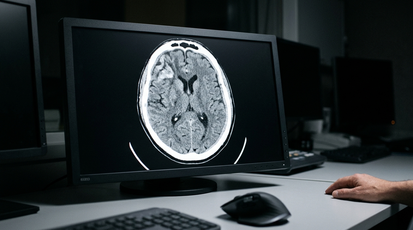
A research team evaluating automated medical imaging techniques has successfully adapted the Segment Anything Model to identify and outline hematomas caused by traumatic brain injury on non-contrast computed tomography scans. By incorporating lightweight adapter modules into the foundational vision transformer architecture, the investigators achieved a slice-level Dice similarity coefficient of 72.34 percent and an intersection over union of 59.78 percent, alongside a ninety-fifth percentile Hausdorff distance of 5.57. The framework demonstrated an overall sensitivity of 75.39 percent and a specificity of 99.73 percent on experimental brain imaging data. Traumatic brain injury represents a profound global health challenge, generating an estimated 50 to 60 million new cases annually. Within acute clinical care, the rapid accumulation of blood clots within brain tissue can cause dangerous escalations in intracranial pressure, secondary neurological deficits, coma, or death, making prompt and accurate volumetric lesion evaluation vital during the immediate golden hours following trauma.
Computed tomography remains the clinical gold standard for acute traumatic brain injury diagnosis and emergency management, yet manual slice-by-slice contouring by expert radiologists remains labor-intensive, time-consuming, and susceptible to substantial inter-observer variability. Automated segmentation methods have increasingly relied on deep learning systems, particularly convolutional neural networks like U-Net and its multi-scale variants. While foundational computer vision models such as the Segment Anything Model offer impressive zero-shot generalization capabilities across diverse visual domains, applying them directly to clinical radiology presents steep obstacles due to high computational overhead and the scarcity of vast annotated medical collections. The vanilla Segment Anything Model yielded poor baseline performance when applied without modifications to traumatic brain injury scans. To overcome these constraints without full parameter retraining, the authors introduced parameter-efficient transfer learning, freezing the primary weights of the base model while training compact bottleneck adapter blocks designed to capture complex intracranial hematoma boundaries.
The study developed a standardized data preprocessing workflow to convert raw Digital Imaging and Communication in Medicine files into standardized volumetric arrays. Axial non-contrast computed tomography slices were converted from raw greyscale values into physical Hounsfield units using linear transformations derived from rescale slope and intercept metadata. While standard clinical cranial windowing frequently relies on a range between 0 and 80 Hounsfield units, traumatic hematoma pathology presents with hyperdense characteristics that appear brighter than normal parenchyma. The researchers therefore established a baseline intensity adjustment window between 0 and 140 Hounsfield units, remapping tissue intensities to enhance pathological visibility. Following intensity rescaling, affine transformations based on calculated anatomical angles ensured uniform three-dimensional brain alignment across cases. Slices were standardized to 512 by 512 pixel grids by applying margin zero-padding to smaller images and center cropping to slices exceeding the standard dimension before network input.
The architectural framework utilizes the Vision Transformer ViT-B base backbone pre-trained with masked auto-encoders, which partitions input images into sixteen-by-sixteen fixed-size patches before routing embeddings through transformer encoder layers containing four global attention blocks. The researchers focused on a sparse prompt encoder configuration operating on point and bounding-box coordinates, followed by a transformer mask decoder featuring two-way cross-attention and a dynamic prediction head. To adapt this system efficiently, serial adapter modules were inserted immediately following the multi-layer attention and multi-layer perceptron sub-layers in each transformer block. Each adapter employs a bottleneck structure, applying a projection matrix that compresses feature dimensionality down to a smaller bottleneck size, followed by a rectified linear unit activation, and an up-projection matrix that restores the original feature dimension. This parameter-efficient structure allowed the team to fine-tune fewer than 4 million adapter parameters while leaving the remaining 86 million base parameters frozen.
The experimental dataset curated for model training and evaluation comprised 119 brain computed tomography scans collected from two distinct clinical sources. A significant portion was drawn from the Progesterone for Traumatic Brain Injury, Experimental Clinical Treatment clinical trial conducted across 49 trauma centers throughout the United States, which enrolled adult patients presenting with moderate to severe head injury and Glasgow Coma Scale scores ranging from 4 to 12. Additional imaging was contributed by the University of Michigan General Neurology Clinic and Emergency Department. Within this primary cohort, 94 scans exhibited confirmed hematomas while 25 scans served as negative controls. All scans were annotated at the voxel level by a board-certified neurologist specializing in neurocritical care, termed Reviewer A. A double-reviewed subset was independently annotated by a second neurocritical care neurologist, Reviewer B, without mutual consultation or visibility into Reviewer A’s contours.
Model training and hyperparameter optimization were executed on NVIDIA Tesla V100 and NVIDIA A40 graphics processing units using the AdamW optimizer with a learning rate of 5e-5, weight decay of 0.1, a batch size of 2, and a 50-epoch schedule across a five-fold cross-validation scheme holding out twenty percent of data for testing. Extensive experiments explored loss function configurations, bottleneck dimensions, and external pretraining. The combination of 50 percent binary cross-entropy and 50 percent boundary loss achieved superior performance over standalone binary cross-entropy, standard Dice loss, and Focal Tversky loss. While Focal Tversky loss achieved strong isolated scores, it induced severe gradient explosions early in training because of volatile fluctuations in the Tversky index weightings. Architectural ablation established that an adapter bottleneck dimension of M equals 100 struck an optimal balance between parameter compactness and segmentation precision, outperforming both smaller and larger bottleneck allocations.
Pretraining experiments tested whether initializing weights on public hemorrhage collections—specifically the Brain Hemorrhage Segmentation Dataset of 192 scans, the INSTANCE 2022 challenge dataset of 100 scans, and the PhysioNet challenge dataset of 82 scans—could boost accuracy. While public pretraining improved baseline representations when input scans lacked contrast adjustment, models trained directly on properly contrast-adjusted windowed slices consistently outperformed those utilizing unadjusted pretraining pipelines. The authors noted that while traumatic and spontaneous hemorrhages share identical hyperdense Hounsfield values, their distinct anatomical distributions and morphology limit transferability. In comparative benchmarking against established architectures, the standard U-Net yielded a Dice score of 56.40 percent and an intersection over union of 44.00 percent, while the Multi-view convolutional network achieved 56.60 percent Dice and 44.40 percent intersection over union. Both convolutional models suffered from low sensitivity, reflecting elevated false negative rates across challenging lesion margins.
The 3D full-resolution nnU-Net benchmark outperformed previous convolutional architectures, demonstrating an 88.03 percent sensitivity, a 65.02 percent Dice score, a 62.76 percent intersection over union, and a 95% Hausdorff distance of 5.89. However, the proposed SAM-Adapter surpassed all tested benchmarks, reaching a 72.34 percent slice-level Dice coefficient and a reduced 95% Hausdorff distance of 5.73. On scan-level volumetric evaluations, the adapted model sustained strong performance, attaining a 75.51 percent Dice coefficient and a 92.05 percent volumetric similarity score. Statistical evaluation via non-parametric Friedman testing across scan-level metrics confirmed significant differences between the models with p-values below 0.05 for Dice, intersection over union, Hausdorff distance, and volumetric similarity. Subsequent post-hoc Nemenyi pairwise comparisons showed no statistically significant performance difference between U-Net and Multi-view CNN, but verified significant stepped performance gains from nnU-Net and the SAM-Adapter architecture.
To evaluate clinical relevance, the investigators examined inter-observer variability between human clinicians and compared it directly against model predictions on the double-reviewed imaging subset. Model predictions compared to human annotations yielded a Dice score of 67.20 percent, whereas direct agreement between the two human reviewers yielded a Dice score of 63.79 percent, an overall accuracy of 95.90 percent, a sensitivity of 59.70 percent, a specificity of 96.31 percent, and a 95% Hausdorff distance of 4.70. Subgroup analysis based on lesion pixel count demonstrated that human agreement peaked on medium-sized hematomas spanning 2,000 to 6,000 pixels. Agreement scores dropped noticeably on very small or very large lesions, with larger lesions generating greater absolute volumetric discrepancies between reviewers.
External validation on the HemSeg-200 dataset, comprising 222 patient computed tomography scans sourced from the Radiological Society of North America with voxel-level intraparenchymal and intraventricular hemorrhage annotations, further confirmed model generalizability. The SAM-Adapter achieved the highest overall segmentation performance, posting an 81.10 percent Dice score and a 70.35 percent intersection over union, outperforming both U-Net and nnU-Net. The authors acknowledged current limitations, noting that their present implementation operates in two dimensions, which restricts spatial continuity relative to fully volumetric 3D architectures like nnU-Net that achieved tighter Hausdorff distances on the external cohort.
Subject of Research: Automated computed tomography hematoma segmentation for traumatic brain injury using an adapted Segment Anything Model
Article Title: Adapting segment anything model for hematoma segmentation in traumatic brain injury
Article References: Cai, L., Williamson, C., Nguyen, A., Wittrup, E., & Najarian, K. (2025). Adapting segment anything model for hematoma segmentation in traumatic brain injury. Discover Imaging, 2(1), Article 6. https://doi.org/10.1007/s44352-025-00011-4
Image Credits: AI Generated
DOI: 10.1007/s44352-025-00011-4
Keywords: Segment Anything Model, traumatic brain injury, hematoma segmentation, computed tomography, parameter-efficient transfer learning, adapter tuning, Hounsfield units, PROTECT III trial, HemSeg-200 dataset, Dice similarity coefficient, Hausdorff distance, nnU-Net benchmark, vision transformer, boundary loss, inter-observer variability, intracranial hemorrhage, binary cross entropy
News Source: Cassandra Pierce. (November 14, 2026). Adapted Segment Anything Model Improves Traumatic Brain Injury Hematoma Segmentation Accuracy. Scienmag.