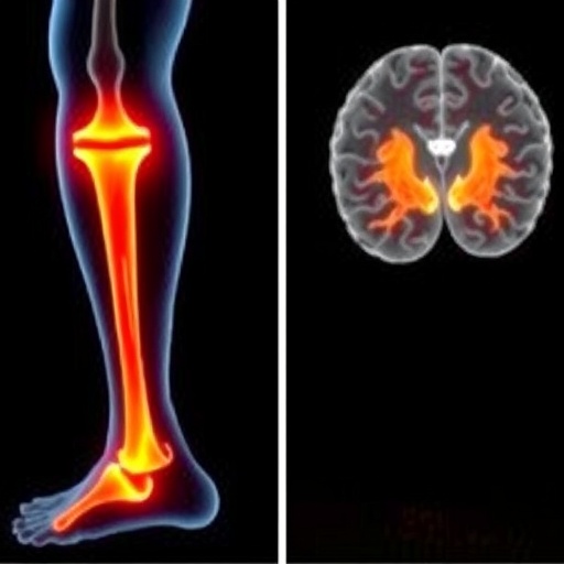
Anterior cruciate ligament injuries may change more than the knee itself. A new scoping review published in Sports Medicine – Open synthesizes evidence from 25 neuroimaging studies and concludes that people with ACL injuries rely on a fundamentally different pattern of brain activity when they move — one that leans heavily on cognitive control and visual processing regions, apparently in compensation for the loss of proprioceptive input from the damaged ligament. The findings strengthen a growing argument that ACL injury should be treated as a neuromusculoskeletal condition, not simply a mechanical joint problem.
The review was led by Robbe Capelleman and Emile Cloet of Ghent University and KU Leuven, together with colleagues at the University of Paderborn, and was conducted according to Joanna Briggs Institute methodological guidance and reported under the PRISMA extension for scoping reviews. The team searched PubMed, Embase, SPORTDiscus and Web of Science from inception to December 31, 2025, and screened 233 records, ultimately including 25 primary studies published between 2008 and 2025. Fifteen of those studies used electroencephalography (EEG), eight used functional magnetic resonance imaging (fMRI), and two used functional near-infrared spectroscopy (fNIRS), together assessing a total of 416 ACL-injured patients at study level. Crucially, unlike earlier reviews that pooled resting-state recordings and sensory-evoked potentials, the authors restricted eligibility to studies measuring brain activity during voluntary motor tasks, so the synthesis speaks more directly to how movement itself is controlled after injury.
The clinical backdrop is sobering. Even after surgical reconstruction and nine to twelve months of criteria-based rehabilitation, only about half of patients return to their preinjury level of sport, up to twenty percent sustain a new ACL injury, and the risk of post-traumatic osteoarthritis remains roughly fourfold elevated. These poor outcomes have long been attributed to persistent alterations in movement quality — stiff landings, reduced knee loading, aberrant cutting mechanics — that linger months to years after return to sport. But traditional rehabilitation addresses these biomechanical impairments almost exclusively from a musculoskeletal perspective. The new review adds weight to the idea that part of the problem lies upstream, in how the central nervous system has reorganized its control strategy.
The rationale is rooted in the biology of the ligament itself. Beyond its role as a mechanical stabilizer, the ACL is packed with mechanoreceptors that transduce proprioceptive information to the central nervous system to support dynamic joint stability. Surgical reconstruction restores mechanical stability but does not replace those native mechanoreceptors, leaving the brain to cope with disrupted afferent signaling from the knee. Under contemporary models of motor control, the brain continuously compares actual sensory input with predicted feedback from its motor commands — so-called forward modeling — and uses the resulting sensory prediction errors to fine-tune movement. When the sensory stream from a joint becomes unreliable, the nervous system is thought to engage sensory reweighting, dynamically adjusting the relative contribution of visual, vestibular and somatosensory inputs to preserve control.
The pattern of results across the included studies fits that framework remarkably well. Twenty-one of the 25 studies reported significant between-group differences in brain activity, and the most consistent finding was heightened engagement of frontal cognitive regions. Across EEG studies, ACL patients showed greater frontal theta power (4–8 Hz) during joint position sense tasks, force reproduction, single-leg stance and dynamic movements such as walking, running, landing and ball kicking. Frontal theta is commonly interpreted as a neural marker of focused attention, error detection and goal-directed decision-making, and is thought to arise in part from the anterior cingulate cortex. Complementary fMRI results echoed this, revealing increased blood-oxygen-level-dependent (BOLD) responses in the frontal gyri, anterior cingulate cortex, inferior frontal pole and paracingulate cortex even during simple knee flexion–extension. The authors interpret this convergence as evidence that ACL patients increasingly depend on conscious, attention-dependent strategies to execute motor tasks that healthy athletes control more automatically.
Visual processing tells a parallel story. Several fMRI studies demonstrated increased BOLD signal in visuospatial regions — the lingual gyrus, lateral occipital cortex and intracalcarine cortex — during isolated knee movements in reconstructed patients, and one study of ACL-deficient patients found greater activation of the posterior inferior temporal gyrus. EEG evidence here was more mixed, with only one study reporting greater parieto-occipital alpha-2 synchronization; however, another found stronger functional connectivity between parieto-occipital and motor regions, suggesting an upregulated visual-motor network supporting balance even when overt performance looked normal. Taken together, the findings tentatively support increased reliance on visual information during movement — a plausible compensation for degraded proprioceptive input, though the authors caution that the pattern appears to vary with task demands.
In contrast, findings for the motor and parietal cortices were markedly heterogeneous. fMRI studies variously reported increased, decreased or unchanged activation of the primary motor cortex, and parietal results diverged both within and across imaging modalities. Some studies showed greater engagement of somatosensory and multisensory integration areas — the secondary somatosensory cortex, precuneus and superior parietal lobule — during isolated knee movements, while others, including both fNIRS studies, found reduced primary somatosensory activation. The authors suggest this heterogeneity may reflect the parietal cortex’s multifaceted role in somatosensory processing, sensorimotor integration, spatial attention and executive function, as well as cortical down-weighting of unreliable somatosensory signals following deafferentation, pain and swelling. Notably, patients with chronic ankle instability show comparable patterns of frontal and visual activation, hinting that peripheral ligament injuries in general may reshape cortical motor control.
The review also examined whether these neural differences actually matter for performance — and here the evidence is more equivocal. Fourteen studies reported behavioral outcomes alongside neuroimaging, and of these, ten found no significant between-group differences in motor performance while four reported impairments. Force reproduction errors, joint repositioning errors, sway velocity, kicking accuracy and countermovement jump performance were mostly preserved despite distinctly different cortical activation. This suggests the alternative neural strategy is largely effective for basic tasks — but not infallible. One study documented greater joint position error despite elevated frontal theta, and another found reduced gamma-band coherence between the motor cortex and quadriceps muscles alongside reduced force steadiness, pointing to impaired cortical drive to the muscle. Interestingly, when attentional demands shifted externally, performance often faltered: three of the four studies employing tasks with an external focus of attention reported worse motor or cognitive performance in ACL patients. The authors frame this as heightened vulnerability to cognitive–motor interference — the decline in performance when cognitive and motor systems compete for the same limited neural resources.
The implications for rehabilitation are potentially significant. Because ACL patients appear to consume more neurocognitive capacity simply to move, standard rehabilitation may under-prepare them for the chaotic, attention-splitting demands of sport. The authors recommend incorporating exercises with an external focus of attention, which on a neural level promote unconscious, automated control processes and free up cognitive resources. They also endorse the “control-to-chaos” framework, in which rehabilitation progresses from controlled settings to increasingly unpredictable environments by adding visual cognitive dual-tasks — such as Stroop tasks — contextual interference, mechanical perturbations or stroboscopic glasses. Such training may increase ecological validity, better simulate game-like situations, and potentially reduce reliance on compensatory cognitive and visual processing. The authors stress, however, that no study has yet tested whether these dual-task interventions actually normalize cortical activation patterns or improve outcomes in ACL-reconstructed patients.
The review is candid about its limitations. All included studies were case-control designs rated as level 3b evidence, with small sample sizes averaging roughly 17 patients per study, considerable heterogeneity in time since injury (ranging from six weeks to over five years), and substantial variability in analysis techniques — from power spectral density and corticomuscular coherence to entropy and phase-lag connectivity measures. Many fMRI paradigms were performed supine with no behavioral data, limiting ecological validity, and eleven of the 25 studies reported no motor performance metrics at all. The authors call for methodological standardization, mobile EEG in realistic environments, longitudinal designs tracking neural adaptations over time, and studies pairing dual-task paradigms with systematic measurement of both motor and cognitive performance. Until then, the link between cortical reorganization and real-world athletic outcomes — including reinjury risk — remains an educated hypothesis. But the direction of the evidence is clear: an ACL rupture rewrites, at least temporarily, the brain’s manual for movement.
Subject of Research: People
Subject of Research: Medicine
Article Title: ACL Injury Alters Brain’s Movement Control, Scoping Review Finds
Article References: Capelleman, R., Cloet, E., Pieters, D., Vanrenterghem, J., Wezenbeek, E., Witvrouw, E., & Lehmann, T. (2026). Distinct Movement-Related Cortical Control Following Anterior Cruciate Ligament Injury: A Scoping Review. Sports Medicine – Open, 12(1), Article 105. https://doi.org/10.1186/s40798-026-01071-4
Image Credits: AI Generated
DOI: 10.1186/s40798-026-01071-4
Keywords: ACL injury brain activity, brain reorganization after ligament injury, cognitive control in knee injury recovery, EEG and fMRI in sports injury research, evidence-based review of neuroimaging in ACL injuries, impact of ACL injuries on motor cortex function, neural mechanisms of movement control post-ACL, neuroimaging findings in ACL injuries, neuromusculoskeletal approach to ACL injury, neuroplasticity in sports-related injuries, proprioceptive deficits and brain changes, visual processing in ACL injury patients
Cite Scienmag News
APA
MLA
Chicago
Cassandra Pierce. (September 8, 2026). ACL Injury Alters Brain’s Movement Control, Scoping Review Finds. Scienmag. https://scienmag.com/acl-injury-alters-brains-movement-control-scoping-review-finds/
Cassandra Pierce. “ACL Injury Alters Brain’s Movement Control, Scoping Review Finds.” Scienmag, 8 September 2026, https://scienmag.com/acl-injury-alters-brains-movement-control-scoping-review-finds/. Accessed 8 September 2026.
Cassandra Pierce. “ACL Injury Alters Brain’s Movement Control, Scoping Review Finds.” Scienmag. September 8, 2026. https://scienmag.com/acl-injury-alters-brains-movement-control-scoping-review-finds/
Copy citation
Download RIS
Tags: ACL injury brain activityACL injury brain activity changesbrain mechanisms of injury compensationbrain reorganization after ligament injurycognitive control in injury compensationcognitive control in knee injury recoveryEEG and fMRI in sports injury researchEEG and fMRI studies in ACL patientseffects of ACL injury on motor controlevidence-based review of neuroimaging in ACL injuriesimpact of ACL injuries on motor cortex functionimpact of ACL injury on movement controlneural mechanisms of movement control post-ACLneuroimaging evidence in sports medicineneuroimaging findings in ACL injuriesneuroimaging in ACL injuriesneuromusculoskeletal approach to ACL injuryneuromusculoskeletal approach to ACL treatmentneuroplasticity after ligament injuryneuroplasticity in sports-related injuriesproprioceptive deficits and brain changesproprioceptive loss and brain adaptationvisual processing in ACL injuryvisual processing in ACL injury patients