Development of new technology for manufacturing a cell-covered material for use in the field of medicine. Embedding treatment cells in implantable medical devices to achieve an increased therapeutic effect and the suppression of adverse effects
Medical materials that can be inserted into the human body have been used for decades in the field of regenerative medicine – for example, stents that can help dilate clogged blood vessels and implants that can replace teeth or bones. The prolonged use of these materials can result in serious adverse effects and loss of various functions – for example, inflammatory responses, generation of fibrous tissues around the material, and generation of blood clots that block blood vessels.
Recently, a Korean research team has drawn attention for developing a technology to reduce the adverse effects by accumulating the peripheral substances of cells on the surfaces of the materials. The Korea Institute of Science and Technology (KIST) has released an announcement that the research team of Dr. Yoon Ki Joung, from the Center for Biomaterials, has successfully developed a material that can be used to accumulate substances present at the cell periphery on the surface of implantable medical materials. The research was carried out in collaboration with the research team of Professor Dong Keun Han, working at the Cha University (President Dong-Ik Kim). This material can be used to deliver cell-based therapeutics to the desired sites as it can be loaded with therapeutic cells such as stem cells.
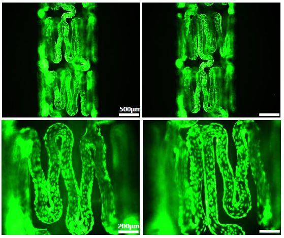
The researchers coated the surface of the material with a compound (polydopamine) and a protein (fibronectin) that can strongly bond with the surface of the material and biomaterial under study. They also used the material surfaces for cell culture studies. The cultured cells produced constituents of the cell periphery (three extracellular matrices). Following this, only the cell was removed and the extracellular matrix was kept intact. This resulted in the generation of space for the attachment of cells necessary for medical purposes. The extracellular matrix enables the adhesion and survival of cells in-vivo because of its high affinity toward cells. Hence, it can effectively deliver the required cells to the treatment sites and mitigate the adverse effects caused by the medical materials.
The researchers applied the developed material to the surface of a stent, a medical device used during surgical procedures to dilate clogged blood vessels. Stents can potentially block blood vessels and cause inflammation or blood clots as they are used to physically extend the blood vessels. This can wound the site that is being operated on. When the developed material was loaded and delivered with endothelial progenitor cells that can regenerate blood vessels, it exhibited excellent vasodilation effects. The damaged inner walls of the blood vessels could be regenerated as well. Regeneration of the inner walls of the blood vessels resulted in a decreased rate (by >70%) of neointimal formation.
Dr. Yoon Ki Joung of KIST said, “This technology can be used to improve various materials that are inserted into the human body. Therefore, it is expected to provide a universal platform for the development of implantable diagnostic and treatment devices (that can potentially dictate the future of technology in the field) and medical devices such as stents and implants that require long-term implantation.”
###
This study was supported by a KIST’s institutional R&D project and the project was supported by the Korea Research Foundation’s Biomedical Technology Development Project, Experienced Researcher Support Project, and Full-cycle medical device R&D Project of All Ministries. It was also supported by the Ministry of Science and ICT (MSIT). The results have been published in the latest issue of “Advanced Functional Materials” (IF: 16.836, top 3.98% in JCR; field: materials science), an international journal.
Media Contact
Do-Hyun Kim
[email protected]
Related Journal Article
http://dx.


