Credit: Associate Professor Takumi Higaki
A research group from Kumamoto University, Japan has developed a highly sensitive technique to quantitatively evaluate the extent of cytoskeleton bundling from microscopic images. Until now, analysis of cytoskeleton organization was generally made by manually checking microscopic images. The new method uses microscopic image analysis techniques to automatically measure cytoskeleton organization. The researchers expect it to dramatically improve our understanding of various cellular phenomena related to cytoskeleton bundling.
The cytoskeleton is a fibrous structure inside the cell made of proteins. It forms higher-order structures called networks and bundles which maintain or change the shape of the cell depending on its state. An accurate understanding of the structures woven by the cytoskeleton makes it possible to estimate the state of a cell. In the past, analysis of the higher-order cytoskeleton structures was generally done by visual observation of the stained cytoskeleton under a microscope by an expert. However, these conventional methods are based on the subjective judgment of the researcher, and thus lack objectivity. In addition, as the number of specimens to be analyzed increases, the personnel costs for experts also grows.
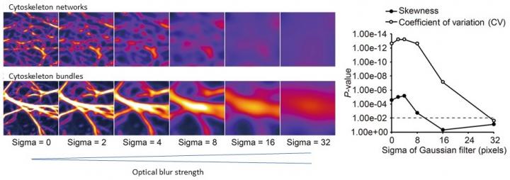
To counter these issues, Associate Professor Takumi Higaki of Kumamoto University has been developing a quantitative method to automatically evaluate the characteristics of complex cytoskeleton structures using microscope image analysis technology. About 10 years ago, he reported that the degree of cytoskeleton bundling could be evaluated by a numerical index he called the “skewness of intensity distribution” from fluorescently stained microscopic images of the cytoskeleton. This technique is now widely used but it has a problem; bundle conditions cannot be accurately evaluated in excessive bundling or when the microscopic image contains a lot of optical blur.
Therefore, Dr. Higaki and a new collaborative research group developed a new quantitative evaluation technique for cytoskeletal bundles that is both more sensitive and versatile than any of the methods described above. Through graphical computer simulations of cytoskeletal bundling, they found that the coefficient of variation of intensities in cytoskeleton pixels nicely reflected the bundle state. Using cytoskeleton microscopic images, they performed a comparative analysis between existing methods and the new method, and found that the proposed method was more sensitive at detecting bundle states than the other methods. They also found that it could be applied to a multitude of biological samples and microscopes. Furthermore, they studied the effect of the proposed method on optical blur–a major cause of image degradation in microscope images–and found that it was sufficient for quantitative evaluation of the bundle state even in unclear images.
“This technology will enable the quantitative evaluation of the state of cytoskeletal bundles from a more diverse set of microscopic images. We expect that it will dramatically advance our understanding of cells by furthering our understanding of higher-order structures of the cytoskeleton,” said Dr. Higaki. “Since cytoskeletal bundling can now be accurately measured, even from unclear images acquired with inexpensive microscopic equipment, new insights may be obtained by reanalyzing the vast amount of microscopic image data that had not been fully utilized in the past.”
###
This research was posted online in Scientific Reports on 21 December 2020.
Source:
Higaki, T., Akita, K., & Katoh, K. (2020). Coefficient of variation as an image-intensity metric for cytoskeleton bundling. Scientific Reports, 10(1). doi:10.1038/s41598-020-79136-x
Media Contact
J. Sanderson & N. Fukuda
[email protected]
Original Source
https:/
Related Journal Article
http://dx.


