A result of NexTEP: Joint Industry-Academia Practical Application Development Project

Credit: YOKOGAWA ELECTRIC CORPORATION
Single-cell mass spectroscopy(1) is a technology to analyze target cell organelles(2) almost as they are, by taking them out from a living cell at a desired time point using a Nanospray Tip(3) while tracking the cell movement under microscope. This technology is highly expected to be a powerful tool to unravel molecular mechanism of the deceases, single-cell diagnosis and personalized medicine.(4) However, existing technology requires manual suction from an individual cell one by one, thus its precision highly depends on the proficiency level of operators. Also, it requires a significantly long time to analyze many cells. Accordingly, demands for automatic suction and much higher sampling efficiency have been growing.
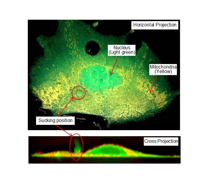
To overcome such problems, we have developed an automatic suction device by combining such technologies as cellular image analysis to select the target, precision positioning of the target location in a cell, and a software for one-click suction. Specifically, you capture microscopy images of cells and calculate their feature quantities, such as the shape and size, and fluorescent intensity. Since such feature quantities reflect the changes caused by drug dosages, they are useful as the objective reference to specify target cells and to maintain high reproducibility of sucked samples. (Fig.1)
Moreover, Yokogawa Electric Corporation’s proprietary confocal microscopy technology(5) and image analysis methods enable three-dimensional measurement of the cell shape and precision positioning of the sucking location at the accuracy of the micron order.
Concerning the detection of the tip position, we have newly developed “Detection by image intensity” method(6) capable for detecting the tip location at the accuracy of less than one micron. (Fig.2 and Fig.3)
Furthermore, learning from the expert operators’ experiences, we have defined the most suitable position for sucking and have incorporated such information in the software, which provides not only the sucking location but also sucking conditions such as pressure and time. As a result, users can suck the target cell organelles by one-click while observing the images of the objective cell. The prototype model, which has all such new features, has successfully obtained a total of 22 samples per hour, which is 4 times more efficient than a manual operation, and thus succeeded in obtaining the specific substances from the target position of the objective cell at high efficiency and precision.
Realization of highly reproducible automatic suction device leads to the practical use of Single-cell mass spectroscopy, which will be widely used for drug discovery, personalized medicine, and so on. In addition to mass spectroscopy, this suction device may be applied for other research, such as sucking a whole cell for mRNA analysis, injection of sucked substances into another cell, etc., and thus will contribute to a wide variety of research fields.
(1) Single-cell mass spectroscopy
A technology to analyze target cell organelles, by trapping and sucking them out from a living cell at a desired time point using a specified thin tube called Nanospray Tip while tracking the cell movement under microscope. Around hundreds femtoliter (fL) sample will be sucked out and analyzed by mass spectroscopy to detect thousands of molecular peaks. In comparison to the conventional analysis based on the mean value of multiple cells, this technology allows for a comprehensive analysis of molecular changes at a single cell level, thus expected to be useful for the unraveling of molecular mechanism of the deceases, single-cell diagnosis, and personalized medicine.
(2) Cell organelles
A general term of intracellular structures, which includes the nucleus to preserve and transfer genetic codes, mitochondria to produce energy, chloroplast to run photosynthesis, etc.
(3) Nanospray Tip
Special thin tube with a tip whose inner diameter is around φ3μm, and outer diameter is around φ5μm. It works as an electrode thus its outer surface is partially coated with conductive materials.
(4) Personalized medicine
Medical treatment considering individual variation of each patient and select drugs and therapy based on the genetic information and symptom of each patient. Also called tailored medicine or made-to-order medicine.
(5) Confocal microscope technology
A microscopy design in which, when the light emitted from a light source focus on a specimen, its reflected light will be focused on the detector surface. Since unfocused light do not reach at the detector surface, only the focused part can be observed significantly brighter. It gives high-resolution and low-noise images, thus allow for highly reliable image analysis. Moreover, it is possible to measure three-dimensional shape of object by capturing and reconstructing images of multiple focal planes.
(6) Detection by Image Intensity Method
A method to detect the location of a sucking tip based on the principle that the tip image becomes brightest when located at the focal point of the objective lens, when moving the tip towards the objective lens under microscope. Since a confocal microscope is used for this device to attain excellent spatial resolution, it is possible to detect the tip at the accuracy level of less than 1μm.
###
Media Contact
Toshiya Ohtake
[email protected]
81-352-148-995
Original Source
http://www.


