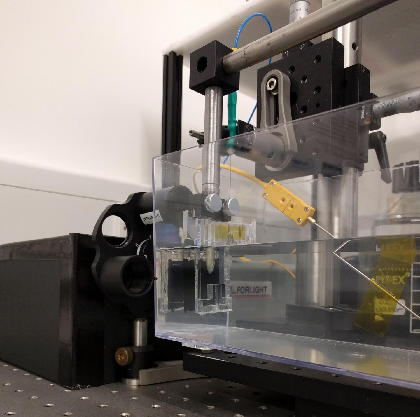

Credit: Erwin J. Alles, University College London
WASHINGTON — While ultrasound is one of the most common medical imaging tools, conventional electronic ultrasound devices tend to be bulky and cannot be used at the same time as some other imaging technologies. A new ultrasound system that uses optical, instead of electronic components, could improve performance while giving doctors significantly more flexibility in how they use ultrasound to diagnose and treat medical problems.
In The Optical Society (OSA) journal Biomedical Optics Express, researchers demonstrate for the first time the use of an all-optical ultrasound imager for video-rate, real-time 2D imaging of biological tissue. The achievement is an important step toward making all-optical ultrasound practical for routine clinical use.
Because they require no electronic components in the imaging probe, all-optical ultrasound systems could be safely used at the same time as magnetic resonance imaging (MRI) scanners. This would give doctors a more comprehensive picture of the tissues around an area of interest, such as a tumor or blood vessel.
"All-optical ultrasound imaging probes have the potential to revolutionize image-guided interventions," said Erwin J. Alles, University College London, United Kingdom. "A lack of electronics and the resulting MRI compatibility will allow for true multimodality image guidance, with probes that are potentially just a fraction of the cost of conventional electronic counterparts."
Lightbeam scanning mirrors built into the device increase image quality and make it possible to acquire images in different modes. In a clinical setting, this would allow doctors to rapidly toggle between modes on a single instrument to suit the task at hand. Acquiring different types of images using conventional ultrasound systems typically requires separate specialized probes.
"The flexibility offered by the scanning mirrors will allow for seamless switching between 2D and 3D imaging, as well as a dynamically adjustable trade-off between image resolution and penetration depth, without the need to swap imaging probe," said Alles. "Especially in a minimally invasive interventional setting, swapping imaging probes is highly disruptive, extends procedure times and introduces risks to the patient."
Eliminating electronics
Conventional ultrasound imagers use arrays of electronic transducers to transmit high-frequency sound waves into tissue and receive the reflections. A computer then constructs images of the tissue.
By contrast, all-optical ultrasound imagers use light to both transmit and receive ultrasound waves. Pulsed laser light is used to generate ultrasound waves, and scanning mirrors control where the waves are transmitted into the tissue. A fiber optic sensor receives the reflected waves.
The electronic components of conventional ultrasound devices make them difficult to miniaturize for internal use, so most existing ultrasound devices are large, handheld probes that are placed against the skin. While some high-resolution minimally invasive ultrasound probes have been developed, they are too expensive for routine clinical use. Optical components are easily miniaturized and tiny all-optical ultrasound probes would likely be significantly less expensive to manufacture than compact electronic ultrasound systems, researchers say.
Speeding up image processing
To generate images, an all-optical ultrasound system must acquire data from multiple optical source locations, combine them together and then create a visualization that reconstructs the area being imaged.
Researchers have previously demonstrated using all-optical ultrasound to generate high-quality 2D and 3D images, but acquiring the images took hours, making these devices too slow to be used in a clinical setting. The new demonstration is the first to acquire and display images with all-optical ultrasound at video rates.
"Through the combination of a new imaging paradigm, new optical ultrasound generating materials, optimized ultrasound source geometries and a highly sensitive fiber-optic ultrasound detector, we achieved image frame rates that were up to three orders of magnitude faster than the current state-of-the-art," said Alles.
A medical multitool
Optical ultrasound systems are inherently more versatile than their electronic counterparts because they can produce sound at a much larger bandwidth. Alles and colleagues demonstrated how the light source can be manipulated to generate either low frequency ultrasound, which results in greater penetration into the tissue, or high frequency ultrasound, which offers higher resolution images at a shallower depth.
The team tested their prototype system by imaging a deceased zebrafish, as well as a pig artery that they manipulated to emulate the dynamics of pulsing blood. The demonstration showed imaging capabilities comparable to an electronic high-frequency ultrasound system, with a sustained frame rate of 15 Hertz, a dynamic range of 30 decibels, a penetration depth of 6 millimeters and a resolution of 75 by 100 micrometers.
To adapt the technology for clinical use, the researchers are working to develop a long, flexible imaging probe for free-hand operation, as well as miniaturized versions for endoscopic applications.
###
Paper: E.J. Alles, S. Noimark, E. Maneas, E.Z. Zhang, I.P. Parkin, P.C. Beard, A.E. Desjardins, "Video-Rate All-Optical Ultrasound Imaging," Biomed. Opt. Express, 9, 3481-3494 (2018). DOI: 10.1364/BOE.9.003481
About Biomedical Optics Express
Biomedical Optics Express is OSA's principal outlet for serving the biomedical optics community with rapid, open-access, peer-reviewed papers related to optics, photonics and imaging in the life sciences. The journal scope encompasses theoretical modeling and simulations, technology development, and biomedical studies and clinical applications. It is published by The Optical Society and edited by Christoph Hitzenberger, Medical University of Vienna. Biomedical Optics Express is an open-access journal and is available at no cost to readers online at OSA Publishing.
About The Optical Society
Founded in 1916, The Optical Society (OSA) is the leading professional organization for scientists, engineers, students and business leaders who fuel discoveries, shape real-life applications and accelerate achievements in the science of light. Through world-renowned publications, meetings and membership initiatives, OSA provides quality research, inspired interactions and dedicated resources for its extensive global network of optics and photonics experts. For more information, visit osa.org.
Media Contacts:
Rebecca B. Andersen
The Optical Society
[email protected]
+1 202.416.1443
Azalea Coste
The Optical Society
[email protected]
+1 202.416.1435
Media Contact
Azalea Coste
[email protected]
202-416-1435
@opticalsociety
http://www.osa.org
Original Source
http://www.osa.org/en-us/about_osa/newsroom/news_releases/2018/versatile_ultrasound_system_could_transform_how_do http://dx.doi.org/10.1364/BOE.9.003481


