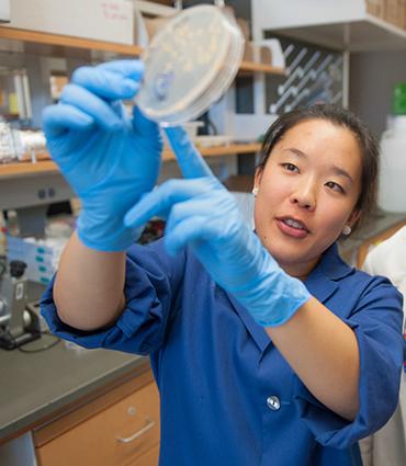

AMHERST, Mass. – Mycobacteria cause a number of dangerous, difficult-to-treat diseases including leprosy and tuberculosis, and progress has been slow in eradicating them. But new strategies for combating these bacteria may eventually emerge from better understanding their basic structure and mechanisms, say molecular microbiologist Yasu Morita and his doctoral student Jennifer Hayashi at the University of Massachusetts Amherst.
In the current issue of Proceedings of the National Academy of Sciences, they report an advance in the fundamental knowledge about a model species of these pathogens, Mycobacterium smegmatis. First author Hayashi and her advisor Morita demonstrate the existence of a distinct domain, or area, on the bacteria's plasma membrane that is crucial for the cell's ability to grow.
Morita says, "This wasn't known and I think many people will be surprised to see that there is such a formal membrane structure there that we didn't expect." The two write that findings provide "an important insight into the potential regulatory mechanisms of lipid metabolism in mycobacteria, where disrupting control points might offer a way to interrupt their growth."
Morita adds, "We hope that discovering this dedicated domain will one day lead to methods of inhibiting bacterial growth, but the real problem of mycobacterial diseases is that these particular bacteria can lie dormant for long periods without active growth. Knowing more about this newly discovered membrane domain could someday let us understand how they control their own growth and how they go dormant to hide in the body."
For this work, Hayashi built on Morita's earlier findings that suggested M. smegmatis's membrane has a specialized domain. Morita says, "I went as far as possible with the techniques available at the time. I showed that this was definitely worth pursuing. I reported a very specific biosynthesis going on in an organized membrane. My data suggested that it's not just a membrane but it's a manufacturing factory of membrane lipids."
Now that techniques such as large-scale comparative proteomics, lipodomics and fluorescence microscopy are available, Hayashi turned to these powerful tools to ask precisely what proteins and lipids, or fats, are present at the membrane domain, and what they do.
She identified more than 300 proteins in the domain and more than 600 in non-domain membrane, a total of nearly 1,000 membrane-associated proteins from a single experiment. "It's a pretty robust approach to figure out what's happening in the domain and non-domain areas, to confirm that the domain is distinct and observe that the two areas have different metabolic activities going on," Hayashi says.
In the lipdomic experiment made possible by collaboration with Branch Moody at Harvard's Brigham and Womens' Hospital, the UMass Amherst microbiologists found more than 600 lipids in the domain and nearly 800 in non-domain regions, providing "another piece of evidence that they are different and have different functions," Hayashi says.
Finally, she fused proteins identified in the proteomics experiment to fluorescent proteins and tracked them to the cell's local domain using fluorescent microscopy. "We were able to note that domain- and non-domain-associated proteins have distinct fluorescent patterns that are maintained while the cell grows. They stay separate, which confirms that this is a spatially distinct domain of the plasma membrane when cells are growing," Morita notes.
The two microbiologists say their discovery is just the latest in the recent series of discoveries showing that the old image of lowly bacteria being simple bags of enzymes is not true.
###
This work was supported by the Mizutani Foundation and the Potts Memorial Foundation, which supports TB research in particular, plus a UMass Amherst Graduate School dissertation grant.
Media Contact
Janet Lathrop
[email protected]
413-545-0444
@umassscience
http://www.umass.edu
The post New structure identified in membrane of disease-causing bacteria appeared first on Scienmag.


