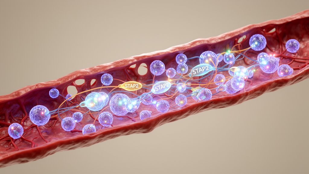
A newly published single-cell transcriptomic investigation in the journal Angiogenesis has uncovered a distinct molecular driver of diabetic retinal vascular injury, identifying signal-transducing adaptor protein 2, or STAP2, as an essential contributor to endothelial dysfunction under diabetic conditions. By combining single-cell RNA sequencing re-analyses of diabetic mouse retinas with comprehensive gain- and loss-of-function experiments in cultured human retinal microvascular endothelial cells and in vivo animal models, the investigators demonstrated that elevated STAP2 expression accelerates microvascular impairment. Mechanistically, the research revealed that STAP2 exerts its deleterious regulatory effects largely through downstream signaling associated with chemokine receptor CXCR4. These findings provide vital clarity into the cellular heterogeneity and molecular cascades governing diabetic vascular complications, offering fresh insights into potential therapeutic targets for preserving inner blood-retina barrier integrity.
Diabetic retinopathy represents one of the most prevalent microvascular complications of chronic diabetes mellitus, remaining a leading cause of acquired vision loss and blindness among working-age adults worldwide. Although current clinical management relies heavily on intravitreal therapies targeting vascular endothelial growth factor, substantial cohorts of patients exhibit incomplete or inconsistent therapeutic responses, underscoring the persistence of alternative pathogenic pathways. However, the precise degree of transcriptional heterogeneity within this specialized vascular compartment and the specific intracellular regulators driving endothelial cells toward pathological states have remained poorly understood due to the technical limitations of conventional bulk tissue profiling techniques.
To overcome these limitations, the research team performed an in-depth bioinformatic re-analysis focused specifically on the endothelial compartment of a single-cell RNA sequencing dataset derived from adult db/db diabetic mice and heterozygous db/m control littermates. After quality control filtering, the full-retina dataset contained 34,010 retinal cells from db/m animals and 51,558 retinal cells from db/db mice. Within this cellular landscape, the investigators utilized established vascular endothelial markers, notably Cdh5 and Pecam1, to isolate 269 high-confidence retinal endothelial cells, encompassing 151 cells from control mice and 118 cells from diabetic mice. Subsequent normalization, principal component analysis across the first twenty-five components, and graph clustering visualized via t-distributed stochastic neighbor embedding resolved three distinct transcriptional subclusters, designated EC1, EC2, and EC3. Crucially, cluster distribution diverged sharply by condition: cluster EC3 was predominantly populated by endothelial cells from diabetic retinas, displaying marked relative upregulation of candidate genes including STAP2, Dock1, Epha4, Adamts1, and Ranbp9.
Comprehensive functional enrichment profiling of the marker genes characterizing the diabetes-associated EC3 subcluster clarified the biological processes activated within these vulnerable endothelial cells. Gene Ontology enrichment evaluations indicated significant overrepresentation of pathways governing the positive regulation of cell migration, apoptotic processes, general transcription factor activity, and GTPase activator activity. Furthermore, Kyoto Encyclopedia of Genes and Genomes pathway mapping linked the EC3 transcriptional program to key intracellular signaling cascades, including Apelin signaling, FoxO signaling, Rap1 signaling, and cellular senescence pathways. Among the differentially expressed transcripts, signal-transducing adaptor protein 2 emerged as a particularly compelling candidate for functional characterization due to its established role as an intracellular scaffold coordinating inflammatory signaling, focal adhesion remodeling, and transcriptional regulation via STAT family proteins and Src-related kinases. Despite documented roles in immune activation, the function of STAP2 in retinal microvascular biology had not been previously evaluated, prompting the hypothesis that STAP2 directly mediates diabetic endothelial dysfunction.
To validate their bioinformatic observations, the authors systematically assessed metabolic phenotypes and retinal tissue parameters in spontaneous leptin receptor-deficient db/db mice alongside age-matched db/m controls at two, four, and six months of age. Metabolic tracking confirmed marked obesity in db/db mice, which weighed an average of 51.17 grams at two months, 61.50 grams at four months, and 66.17 grams at six months, compared to 27.17, 31.67, and 29.83 grams in control groups. Random blood glucose levels were severely elevated in db/db mice, averaging 23.62 millimoles per liter at two months and surpassing the glucometer detection limit of 33.3 millimoles per liter in multiple animals at older ages. To establish broader generalizability, the researchers instituted an independent chemically induced diabetic model using male C57BL/6J mice administered streptozotocin at 55 milligrams per kilogram daily over six consecutive days. Additionally, surgically excised fibrovascular membrane specimens obtained from a patient undergoing vitrectomy for active proliferative diabetic retinopathy provided clinically relevant translational tissue.
Confocal immunofluorescence microscopy of retinal sections and whole mounts confirmed baseline STAP2 localization within CD31-positive retinal vessels of non-diabetic controls and demonstrated a significant escalation of vascular-associated STAP2 immunoreactivity in diabetic retinas. Quantitative image analysis revealed a statistically significant increase in the background-corrected STAP2-to-CD31 mean fluorescence intensity ratio in db/db mice across two, four, and six months of age. Quantitative morphometry of superficial, intermediate, and deep capillary plexuses confirmed that this elevated fluorescence ratio did not reflect differences in overall vascular density, which remained comparable between age-matched groups. Western blot analyses of whole-neural-retina protein lysates independently demonstrated sustained increases in retinal STAP2 abundance in diabetic mice across all examined ages. Complementary immunostaining verified intensified vascular STAP2 signals in streptozotocin-induced diabetic mouse retinas and confirmed STAP2 expression within CD31-positive vessels of human proliferative diabetic retinopathy tissues. Finally, culturing human retinal microvascular endothelial cells under glucose concentrations of 30 and 50 millimoles per liter stimulated substantial increases in STAP2 protein abundance.
To directly interrogate the cellular consequences of altered STAP2 levels, the investigators conducted in vitro gain- and loss-of-function assays using human retinal microvascular endothelial cells transduced with lentiviral vectors. High-glucose conditions containing 30 millimoles per liter glucose elevated baseline endothelial cell proliferation, migratory capacity in twenty-hour Transwell assays, and early tubular capillary morphogenesis on Matrigel matrices evaluated at four hours post-seeding. Targeted genetic silencing of STAP2 using two independent short hairpin RNA constructs significantly suppressed this high-glucose-induced hyperproliferative phenotype and curtailed cell migration. Furthermore, STAP2 knockdown markedly attenuated early tube formation, substantially diminishing total tube length, total segment piece counts, and master branch junctions quantified through automated angiogenesis analysis. Conversely, stable lentiviral overexpression of STAP2 under normal physiological glucose concentrations of 5.5 millimoles per liter was sufficient to enhance endothelial proliferation, accelerate transwell chemotactic migration, and augment network branching complexity, demonstrating that STAP2 acts as a potent cell-autonomous promoter of angiogenic activity in human retinal endothelial cells.
Translating these findings into living organisms, the researchers evaluated the direct pathological impact of retinal STAP2 modulation in vivo using intravitreal administration of adeno-associated virus serotype 2/1 delivery vectors. Four-week-old db/db mice received vectors expressing STAP2-targeting short hairpin RNA driven by a U6 promoter, while normoglycemic C57BL/6J mice were injected with vectors overexpressing full-length mouse STAP2 under a cytomegalovirus promoter. Following retinal harvest at six months of age, vascular preparations isolated via retinal trypsin digestion revealed that diabetic db/db mice exhibited severe microvascular degeneration characterized by high numbers of non-perfused acellular capillaries. Remarkably, viral knockdown of retinal STAP2 significantly preserved capillary architecture, causing a substantial reduction in acellular capillary formation. In parallel, retro-orbital Evans blue dye extravasation assays confirmed that STAP2 depletion significantly curbed pathological retinal vascular permeability in diabetic animals. Conversely, virally driven STAP2 overexpression in healthy non-diabetic mice triggered pronounced acellular capillary dropout and severe vascular leakage, demonstrating that retinal STAP2 elevation actively drives structural vascular decay.
To delineate the molecular pathways operating downstream of STAP2, the authors conducted unbiased transcriptomic profiling using bulk RNA sequencing across two distinct experimental paradigms in cultured human retinal microvascular endothelial cells. Differentially expressed genes identified in cells overexpressing STAP2 under normal glucose conditions were directly intersected with genes downregulated following STAP2 knockdown under high-glucose conditions, yielding a prioritized core set of 75 overlapping responsive transcripts. Gene Ontology analyses of these candidate genes demonstrated substantial enrichment in cell adhesion, apoptotic regulation, directional motility, and membrane protein ectodomain proteolysis. Among these candidates, chemokine receptor CXCR4 was selected for extensive mechanistic validation due to its established involvement in vascular homing, endothelial migration, and pathological neovascularization. Quantitative real-time polymerase chain reaction and Western blotting confirmed that STAP2 overexpression significantly upregulated CXCR4 mRNA and protein abundance, whereas STAP2 silencing diminished CXCR4 levels. Correspondingly, CXCR4 protein expression was elevated in neural retinas from db/db mice and in endothelial cells exposed to prolonged high-glucose stress.
To determine whether CXCR4 signaling is functionally required for STAP2-induced endothelial activation, the investigators performed targeted rescue experiments by transfecting STAP2-overexpressing endothelial cells with small interfering RNA specifically silencing CXCR4. Functional evaluations demonstrated that knocking down CXCR4 significantly attenuated the heightened cellular proliferation, Transwell migration, and Matrigel capillary network formation induced by STAP2 overexpression. While these data establish CXCR4-associated signaling as an essential downstream functional branch, the authors noted several critical experimental limitations, including the modest sample size of 269 endothelial cells within the foundational single-cell dataset, which renders the initial clustering exploratory. Furthermore, because transcriptomic gain- and loss-of-function profiling was performed under differing glucose backgrounds, overlapping candidates represent exploratory associations rather than bidirectional regulation under identical conditions. Additionally, non-endothelial-specific viral promoter targeting in vivo reflects whole-tissue responses, and human diabetic tissue confirmed vascular presence rather than quantitative overabundance relative to healthy controls. Nevertheless, the study provides compelling experimental evidence linking diabetes-associated STAP2 induction and CXCR4 signaling to retinal endothelial dysfunction and microvascular injury.
Subject of Research: Role of STAP2 and CXCR4 signaling in diabetic retinal endothelial dysfunction
Article Title: STAP2 contributes to retinal endothelial dysfunction in diabetes through CXCR4-associated signaling revealed by single-cell RNA sequencing
Article References: Xiao, Y., Zhu, Q., Zhang, J., Lv, K., Wang, H., Liu, X., Jiang, Y., Shi, X., Qu, Y., Xing, X., Liu, X., Niu, T., & Liu, K. (2026). STAP2 contributes to retinal endothelial dysfunction in diabetes through CXCR4-associated signaling revealed by single-cell RNA sequencing. Angiogenesis, 30(1), Article 2. https://doi.org/10.1007/s10456-026-10106-z
Image Credits: AI Generated
DOI: 10.1007/s10456-026-10106-z
Keywords: diabetic retinopathy, STAP2, CXCR4, retinal endothelial cells, single-cell RNA sequencing, microvascular dysfunction, blood-retina barrier, vascular leakage, acellular capillaries, HRMECs, db/db mice, angiogenesis, transcriptomics, streptozotocin, Evans blue extravasation, cell migration, endothelial heterogeneity
News Source: Drew Townsend. (November 22, 2026). Single-Cell Sequencing Uncovers How STAP2 Drives Diabetic Retinal Vascular Dysfunction. Scienmag.