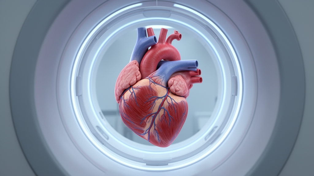
A focused review published in EJNMMI Physics examines how integrating simultaneous magnetic resonance imaging with positron emission tomography can mitigate complex cardiac and respiratory motion to achieve quantifiable, high-resolution cardiac imaging. Producing reliable cardiac PET images requires overcoming independent and highly variable respiratory and heart movements. These physiological motions frequently exceed the thickness of the left ventricular wall, introducing blur that compromises diagnostic precision and the quantification of cardiovascular diseases.
Magnetic resonance imaging currently excels at resolving transmural differences across the left ventricular wall, an anatomical capability that cardiac PET alone has not fully achieved. However, technical advancements in PET spatial resolution are approaching three millimeters. This engineering progress indicates that transmural quantification is feasible if cardiac and respiratory displacements are effectively corrected during data acquisition and image processing.
Simultaneous hybrid PET and MRI platforms provide a synergistic framework by acquiring co-registered multimodal datasets in parallel. Under this approach, the superior anatomical resolution of MRI directly informs PET image reconstruction. This combined architecture permits spatial resolution enhancement and motion correction without incurring the severe signal-to-noise ratio penalties typical of traditional prospective or retrospective gating strategies.
Key motion mitigation strategies highlighted in the review rely on deriving accurate motion vector fields directly from Cine MRI sequences. When investigators apply these MRI-derived motion vectors to acquired PET emission data, the resulting reconstructions display substantial improvements in cardiac image sharpness and overall quality.
Beyond motion vector tracking, integrating anatomical priors from MRI into PET reconstruction algorithms provides additional resolution enhancement, which proves particularly critical when imaging thin myocardial walls. Furthermore, machine learning frameworks offer transformative potential to accelerate these complex correction pipelines, creating operational pathways for demanding applications such as multi-tracer injection protocols.
By overcoming respiratory and cardiac motion artifacts, hybrid PET and MRI systems deliver the quantifiable, high-resolution cardiac data necessary for rigorous preclinical investigations and precise clinical theranostic evaluations in heart disease management.
Subject of Research: Cardiac PET/MRI motion correction strategies and high-resolution quantification
Article Title: Defeating the Black Knight: a focused review of cardiac PET/MRI motion correction
Article References: Lim, H., Wilk, B., Moran, G., Faul, D., Thiessen, J. D., & Prato, F. S. (2026). Defeating the Black Knight: a focused review of cardiac PET/MRI motion correction. EJNMMI Physics. https://doi.org/10.1186/s40658-026-00973-2
Image Credits: AI Generated
DOI: 10.1186/s40658-026-00973-2
Keywords: PET/MRI, cardiac imaging, motion correction, respiratory motion, cardiac motion, left ventricular wall, transmural quantification, image reconstruction, Cine MRI, motion vectors, anatomical priors, machine learning, multi-tracer studies, preclinical research, theranostics, positron emission tomography
News Source: Denise Maddox. (November 20, 2026). Hybrid PET-MRI Motion Correction Enables High-Resolution Cardiac Disease Quantification. Scienmag.