Chronic hypertension drives progressive structural and functional alterations within the cerebral circulation, establishing early pathological pathways that link high blood pressure to blood-brain barrier breakdown, cerebral small vessel disease, and neurovascular decoupling. Synthesizing evidence from specialized rodent models such as spontaneously hypertensive rats and stroke-prone strains, researchers have mapped how sustained mechanical pressure inside cerebral arteries induces inward microvascular remodeling, impairs myogenic autoregulation, and triggers localized cellular inflammation long before clinical cognitive decline or ischemic lesions become apparent.
Clinical metrics establish the diagnosis of hypertension when systolic blood pressure reaches or exceeds 130 mmHg, or diastolic pressure reaches or exceeds 90 mmHg across two separate evaluation days. Worldwide, hypertension impacts approximately 1.13 billion individuals and accounts for roughly 9.4 million deaths annually as a leading contributor to fatal cardiovascular complications. While physiological mechanisms in healthy organisms preserve blood pressure through balanced regulatory networks, chronic disruption of these systems exposes delicate cerebral capillaries to sustained hemodynamic stress, making the brain one of the earliest and most severely affected target organs.
To investigate the intricate pathophysiology of hypertensive cerebrovascular injury, experimental research relies heavily on established in vivo rodent strains. The spontaneously hypertensive rat, initially bred in Kyoto from Wistar ancestors exhibiting elevated blood pressure and stabilized through sibling mating, spontaneously develops elevated blood pressure into adulthood. At six months of age under standard dietary conditions, these animals demonstrate significantly higher systolic blood pressure compared to normotensive Wistar Kyoto controls. Adding dietary salt exacerbates these pressure elevations alongside disturbances in lipid metabolism, allowing scientists to track hypertensive vascular changes over time.
These stroke-prone animals exhibit structural anomalies mirroring human cerebral small vessel disease, including microvascular wall damage, blood-brain barrier leakage, erythrocyte extravasation into brain parenchyma, punctate cerebral hemorrhages, and myelin loss. Furthermore, they display accelerated dysregulation of blood pressure regulatory genes including Agt, Agtrap, Ephx2, and Uts2 compared to baseline hypertensive strains.
Complementary rodent lines provide additional insights into diverse hypertensive mechanisms and systemic interactions. Dahl salt-sensitive rats, developed from selectively bred Sprague-Dawley stock, manifest severe hypertension and cardiac hypertrophy when exposed to high-sodium diets containing between 2% and 8% salt. These animals exhibit impaired pressure-natriuresis, decreased venous compliance, elevated vascular resistance, altered renin-aldosterone dynamics, and progressive renal injury resembling human chronic kidney disease, alongside sodium-induced elevations in plasma PCSK9 that suppress low-density lipoprotein receptors to trigger hypercholesterolemia.
Genetically hypertensive mouse models, particularly the BPH/2J strain established through selective breeding based on tail-cuff systolic blood pressure measurements, illuminate central neurogenic drivers of high blood pressure. In contrast to normotensive BPN/3 controls, BPH/2J mice exhibit marked sympathetic nervous system hyperactivation, elevated circulating adrenaline and noradrenaline, and pronounced c-fos activation within the medial amygdaloid nucleus. Diminished expression of specific GABA-A receptor subunits in the hypothalamus compromises tonic neuronal inhibition, while duplicated orexin mRNA levels further sustain sympathetic tone, showing that central amygdala-hypothalamic pathways can drive systemic vascular constriction.
Within the cerebral vasculature, sustained elevated intraluminal pressure forces arterial walls to undergo structural inward remodeling to counter elevated wall tension and circumferential stress. Morphometric analyses and pressure myography in hypertensive rat models demonstrate significant reductions in arterial lumen diameter accompanied by increased media-to-lumen ratios. In basilar arteries isolated from young hypertensive rats, proteomic analyses demonstrate vascular hypertrophy alongside imbalanced expression of extracellular matrix and angiogenic proteins, confirming that luminal narrowing stems from active structural rearrangement rather than simple wall thickening alone.
Much of this inward remodeling and arterial wall stiffening is driven by angiotensin II signaling rather than elevated mechanical pressure alone. Experimental continuous infusion of angiotensin II in healthy mice induces persistent inward arteriolar remodeling even under moderate blood pressure elevations, mediated in part through caveolin-1 activation pathways. In spontaneously hypertensive rats, chronic arteriolar stiffening elevates basal vasoconstrictive tone in leptomeningeal anastomoses, restricting collateral blood flow during experimental ischemia and reperfusion, while in salt-sensitive rats, central arterial stiffening correlates directly with hippocampal hypoperfusion and reduced N-acetyl aspartate levels indicative of early neuronal stress.
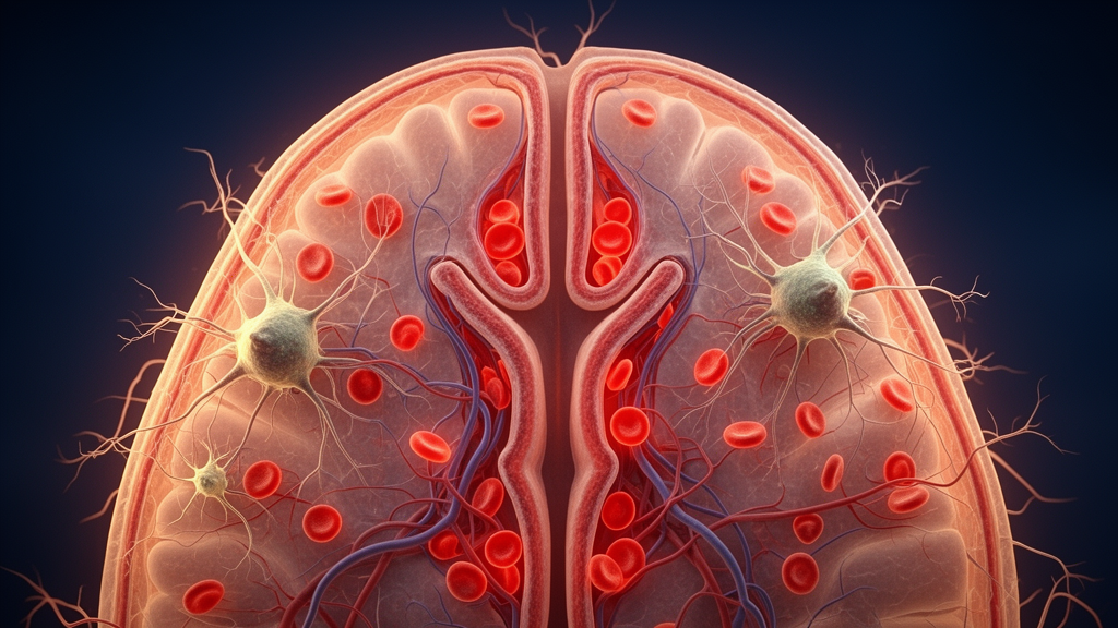
Under normal conditions, cerebral blood flow autoregulation preserves relatively constant microvascular perfusion across fluctuations in mean arterial pressure, but hypertension impairs myogenic contractile responses across standard physiological pressure gradients. Studies demonstrate that sustained hypertension stimulates pericytes to boost perivascular collagen deposition and basement membrane thickening, disrupting crucial endothelial-pericyte signaling interfaces and compromising blood-brain barrier integrity before capillary rarefaction and cognitive deficits emerge.
At the molecular level, blood-brain barrier disruption involves alterations to tight junction architecture and elevated transcytosis mediated by angiotensin II type 1 receptors. Activation of these receptors on perivascular macrophages stimulates the Nox2-containing NADPH oxidase enzyme complex to generate substantial volumes of reactive oxygen species, driving oxidative stress across the neurovascular unit. Longitudinal evaluations confirm that this oxidative burden suppresses cortical hemodynamic coupling during functional sensory stimulation, establishing an early mechanistic bridge whereby chronic hypertensive vascular remodeling leads to subcortical small vessel disease, white matter damage, and impaired cognitive function.
Direct experimental assessments of blood-brain barrier permeability have clarified the cellular timeline of microvascular failure across hypertensive strains. When researchers tracked the transit of intravenous horseradish peroxidase in three-month-old rats, spontaneously hypertensive rats and stroke-prone spontaneously hypertensive rats exhibited marked tracer extravasation from vessels along the hippocampal fissure, whereas normotensive Wistar Kyoto controls retained vascular containment. Ultrastructural imaging of stroke-prone animals located horseradish peroxidase within cytoplasmic endothelial vesicles, the basal lamina, and extracellular spaces, accompanied by perivascular collagen deposition. Under physiological conditions, endothelial cells maintain barrier selectivity through tight junctions that prevent paracellular diffusion alongside specialized luminal transporters including fatty acid transport protein 1 and MFSD2A. However, sustained hypertensive stress disrupts these regulated cellular interactions among endothelium, pericytes, and astrocytes, though therapeutic administration of the angiotensin-converting enzyme inhibitor perindopril inhibited inward arteriolar remodeling in stroke-prone rats.
These microvascular injuries accelerate the presentation of cerebral small vessel disease, which damages subcortical white matter, periventricular white matter, and deep gray nuclei. In clinical populations, hypertension is diagnosed in approximately 70 percent of adults older than 65 years and occurs in 80 percent of patients with cerebral small vessel disease. In contrast, amyloid variants involve the vascular accumulation of amyloid-beta fibrils, known as cerebral amyloid angiopathy, appearing sporadically in over 50 percent of individuals past 80 years of age. Anatomically, type 1 amyloid angiopathy restricts fibril deposition predominantly to cerebral capillaries and microvessels, whereas type 2 angiopathy preferentially targets small intracortical arteries, arterioles, and superficial pial or meningeal vessels.
Rodent investigations have shown that non-amyloid vascular damage and amyloid pathology interact as a continuous spectrum. In stroke-prone spontaneously hypertensive rats, chronic high blood pressure impairs amyloid-beta clearance, generating a microvascular substrate that promotes secondary amyloid accumulation. Longitudinal evaluations combining in vivo two-photon microscopy, localized blood flow recordings, and ex vivo immunohistochemistry for amyloid-beta, immunoglobulin G, and lectins demonstrated that blood-brain barrier breakdown, arteriolar wall injury, and microvascular thrombotic events arise prior to demonstrable amyloid deposition. Under baseline conditions, healthy neurovascular coupling relies on reciprocal signaling among neurons, astrocytes, pericytes, smooth muscle, and endothelium mediated through nitric oxide, prostaglandins, adenosine, and specific ion channel cascades. Neuronal activation stimulates endothelial relaxation at the capillary level, which propagates retrogradely into upstream arterioles to boost localized perfusion; however, hypertensive microvascular damage disrupts this communicative cascade.
The molecular signaling governing neurovascular disruption involves distinct cellular compartments and central endocrine circuits. Within the cerebral microvasculature, myeloid-derived perivascular macrophages express functional angiotensin II type 1 receptors and generate substantial amounts of reactive oxygen species through the Nox2 enzyme complex. Arteriolar smooth muscle cells lack both type 1 receptors and Nox2 expression, while single-cell RNA sequencing indicates that endothelial cells express only modest amounts of Nox2 compared to macrophages and microglia. Consequently, targeted depletion or genetic deletion of perivascular macrophages completely abolishes vascular free radical production during slow-pressor angiotensin II hypertension, whereas acute administration engages endothelial pathways.
Functional neuroimaging studies in spontaneously hypertensive rats further illustrate the persistent nature of neurovascular decoupling. By six months of age, these animals exhibit marked reductions in cortical hemodynamic responses to whisker stimulation despite displaying intact baseline neuronal metabolism. Spectroscopic and optical imaging reveal that while antihypertensive pharmacological treatments normalize baseline cerebral perfusion, they fail to rescue stimulus-evoked functional hyperemia, highlighting enduring vascular dysfunction. Magnetic resonance imaging demonstrates that microvascular hemodynamics transition from early transient hyperperfusion to chronic hypoperfusion and coupling loss, aligning with microvascular rarefaction and neural cell loss. Significant experimental gaps persist because functional coupling remains largely uncharacterized in stroke-prone rats due to early lesion interference, while standardized evaluation protocols remain absent for Dahl salt-sensitive models.
Subject of Research: Rodent models of hypertension-induced cerebrovascular remodeling and brain damage
Article Title: Hypertension and brain damage: evidence from rodent models
Article References: Scenna, M. S., Maceroni, E., Cimini, A., Castelli, V., & d’Angelo, M. (2026). Hypertension and brain damage: evidence from rodent models. Laboratory Animal Research, 42(1), Article 9. https://doi.org/10.1186/s42826-026-00270-0
Image Credits: AI Generated
DOI: 10.1186/s42826-026-00270-0
Keywords: hypertension, cerebrovascular disease, blood-brain barrier, neurovascular coupling, rodent models, spontaneously hypertensive rats, stroke-prone SHR, cerebral autoregulation, microvascular remodeling, angiotensin II, perivascular macrophages, oxidative stress, pericytes, cerebral small vessel disease, arteriolar remodeling
News Source: Cassandra Pierce. (November 19, 2026). Rodent Models Reveal How Chronic Hypertension Triggers Structural Brain Damage. Scienmag.