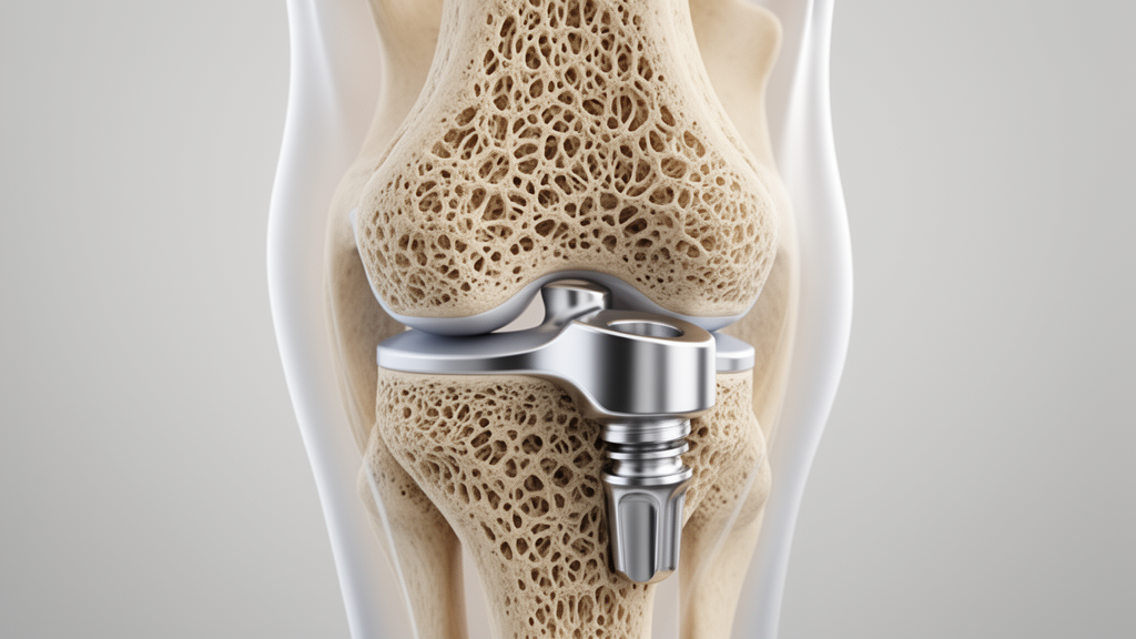
Cementless total knee arthroplasty has experienced a major resurgence in orthopedic surgery, particularly among younger, active, and obese patients facing elevated risks of aseptic loosening with traditional cemented fixation. Modern manufacturing innovations, including additive three-dimensional printing and advanced porous coatings like hydroxyapatite beads and biomimetic trabecular matrices, have significantly heightened clinical expectations for durable biologic osseointegration. Nevertheless, as highlighted in a narrative review in Knee Surgery and Related Research by Dong Hwan Lee and colleagues, these bioengineering advancements cannot guarantee success without adequate initial mechanical stability at the bone-implant interface. Inadequate initial fixation permits excessive micromotion, precipitating fibrous tissue encapsulation rather than osseointegration and culminating in early implant failure. Because initial stability depends fundamentally on the local mechanical competence of host bone, accurate site-specific bone quality assessment has emerged as an indispensable prerequisite for candidate selection in cementless knee reconstruction.
Evaluating bone quality for cementless knee replacement requires a fundamental shift away from traditional systemic osteoporosis diagnostics toward anatomically focused regional measurements. Central dual-energy X-ray absorptiometry measuring the lumbar spine and femoral neck remains the clinical gold standard for diagnosing systemic osteopenia and osteoporosis, conditions frequently underdiagnosed across arthroplasty cohorts. However, mechanical and clinical investigations demonstrate that central bone mineral density correlates weakly with the actual compressive strength of the distal femur and proximal tibia. In receiver operating characteristic curve analyses applying mechanical criteria for cementless suitability, central measurements demonstrated poor diagnostic performance, confirming that systemic values cannot reliably predict regional peripheral support. This discrepancy is magnified by the biomechanical pathology of knee osteoarthritis, where joint malalignment and altered weight-bearing distributions cause asymmetric bone remodeling. In varus osteoarthritic knees, for instance, mechanical overload induces subchondral bone sclerosis in medial condyles, whereas the unloaded lateral compartment experiences marked bone loss.
To address the need for accessible localized screening, researchers have explored plain radiography, the most ubiquitous imaging modality in routine orthopedic practice. Inspired by hip arthroplasty metrics such as the Dorr classification, cortical index, and canal-to-diaphysis ratio, investigators developed knee-specific radiographic indices, including the reverse Dorr concept correlating distal femoral cortical morphology with osteopenia. Other radiographic strategies utilize grayscale densitometric analysis, quantifying regional pixel intensity within predefined regions of interest calibrated against internal references like aluminum step wedges or software platforms such as IBEX Bone Health. Similarly, trabecular bone texture analysis attempts to quantify structural heterogeneity and microarchitectural complexity as surrogate markers of subchondral organization. While standard knee radiographs offer opportunistic screening for low bone mass, they possess severe inherent limitations. Planar two-dimensional projection, rotational positioning variability, and soft-tissue attenuation confound measurements, while cortical indices fail to capture cancellous trabecular strength directly supporting the cut surface, leaving plain radiography unvalidated as a definitive candidate selection tool.
Alternative two-dimensional and cross-sectional imaging modalities have also been investigated to bridge the diagnostic gap between systemic screening and localized surgical planning. Knee-specific peripheral dual-energy X-ray absorptiometry permits targeted evaluation of localized regions of interest across the femoral and tibial condyles, effectively identifying compartmental bone density disparities driven by limb alignment. Nevertheless, peripheral knee absorptiometry continues to suffer from planar two-dimensional projection and lacks standardized acquisition protocols, restricting its role to adjunctive screening. Magnetic resonance imaging provides three-dimensional visualization of marrow composition and trabecular microarchitecture without ionizing radiation. Adapting concepts like the vertebral bone quality score from spine surgery, researchers measured fat-suppressed signal intensity ratios between knee bones and synovial fluid, finding higher ratios in osteoporotic bone. However, this magnetic resonance approach achieved a modest predictive accuracy of sixty-five percent in distal femoral bone, and high-resolution trabecular microarchitecture imaging remains unvalidated against direct mechanical bone strength, restricting magnetic resonance imaging from routine clinical adoption in arthroplasty decision-making.
Computed tomography is currently considered the most practical and clinically powerful modality for knee-specific bone quality assessment, delivering three-dimensional structural data that aligns with routine preoperative workflows and robotic-assisted surgical planning. Conventional computed tomography allows clinicians to measure Hounsfield unit attenuation values across customized regions of interest without requiring specialized software or hardware. Attenuation reflects tissue density standardized against water and air, and opportunistic knee screening studies by Klinger and Sebro demonstrated robust correlations between regional Hounsfield units and systemic bone density. Crucially, recent mechanical investigations directly linked computed tomography attenuation to structural competence, demonstrating significant positive correlations between preoperative Hounsfield units and compressive strength measured during mechanical testing of polyurethane blocks and distal femoral box bone resections. Furthermore, retrospective analyses of robotic arthroplasty cohorts revealed that patients selected for cementless fixation exhibited significantly higher proximal tibial attenuation (152.7 Hounsfield units) than cemented cohorts (105.6 Hounsfield units), highlighting its practical utility despite potential calibration variability across scanner platforms.
To overcome the limitation that conventional Hounsfield unit values provide relative attenuation estimates rather than absolute physical density, advanced computed tomography modalities have been introduced. Quantitative computed tomography utilizes calibrated phantom references—or emerging phantomless algorithms referencing internal muscle and adipose tissue—to convert attenuation values into standardized volumetric bone mineral density in milligrams per cubic centimeter. Although clinically established for spinal osteoporosis and vertebral fracture assessment, its procedural complexity and calibration requirements have limited routine knee application. In contrast, dual-energy computed tomography employs simultaneous acquisitions at two distinct X-ray energy spectra, executing material decomposition to separate mineral components from soft tissues. Investigations by Choi and Lee demonstrated that dual-energy computed tomography volumetric bone mineral density accurately screens for regional osteopenia and correlates strongly with actual distal femoral compressive strength in mechanical testing. These findings confirm that dual-energy computed tomography offers highly accurate volumetric assessment, although widespread clinical adoption still requires protocol standardization and accessible infrastructure.
While preoperative imaging establishes baseline anatomical data, intraoperative assessment provides the irreplaceable advantage of directly evaluating the freshly prepared surgical implant bed. Historically, intraoperative judgment relied entirely on subjective tactile feedback, exemplified by the classic thumb test adapted from shoulder surgery and the bone hardness test employed in unicompartmental knee arthroplasty. In these maneuvers, the surgeon applies manual pressure with the thumb against the resected tibial surface, selecting cemented fixation if deflection occurs or cementless components if the bone resists depression. To introduce greater consistency, investigators have attempted to systematize surgical perception. Nickel and colleagues categorized haptic feedback encountered during bone cutting and drilling, demonstrating meaningful correlations with distal femoral bone mineral density. Similarly, Maniar and co-workers implemented a visual-tactile grading scale ranging from zero to ten that correlated positively with femoral neck absorptiometry. Despite their structured approach, these semi-quantitative frameworks remain fundamentally dependent on individual surgeon sensory interpretation and applied mechanical force.
Recent technological innovations have focused on eliminating subjective tactile bias through objective morphological classification and mechanical measurement devices. Lee and colleagues introduced an intraoperative visual assessment system categorizing distal femoral cut surface morphology into four distinct grades—excellent, good, fair, and poor—based strictly on inspecting trabecular pore characteristics and contour integrity. When validated against mechanical compression testing of resected femoral bone specimens, this visual grading system demonstrated strong correlation with actual compressive strength, achieving outstanding diagnostic accuracy on receiver operating characteristic analysis with an area under the curve of 0.941. In parallel, device-based approaches have utilized torque-measuring probes such as the Densiprobe, which features a wingblade mechanism quantifying peak trabecular torque inside femoral peg holes during unicompartmental procedures. Although providing direct mechanical measurements that correlate with absorptiometry metrics, their utility remains restricted because they cannot evaluate implant designs lacking peg holes, sample only localized entry points rather than the entire resection bed, and lack validation in total knee arthroplasty.
A critical disparity highlighted by the review is that existing objective intraoperative assessment tools have been validated almost exclusively on the distal femoral cut surface, leaving the proximal tibia comparatively unstudied. This research gap stems primarily from methodological constraints: resected proximal tibial bone wafers are generally inadequate for mechanical compression testing, and tibial preparation rarely incorporates deep peg holes suitable for torque probe insertion. Nevertheless, addressing tibial bone quality is paramount because component migration, progressive subsidence, and aseptic loosening occur far more frequently on the tibial side in cementless total knee arthroplasty. Tibial evaluation is especially challenging in varus osteoarthritic knees, where dense subchondral sclerosis in the diseased medial compartment can deceive surgeons into overestimating overall tibial bed quality. Underneath this sclerotic surface, adjacent metaphyseal cancellous bone and the unloaded lateral compartment often suffer severe structural compromise, highlighting the necessity for objective validation protocols applied directly to the proximal tibial resection bed.
Looking toward the future, candidate selection for cementless total knee arthroplasty is expected to advance through artificial intelligence integration, multi-modal diagnostic modeling, and perioperative metabolic optimization. Machine learning and deep learning architectures hold potential to automate three-dimensional region of interest segmentation, attenuation mapping, and fracture risk stratification from standard computed tomography and radiographic data. Future protocols will likely evaluate site-specific cancellous strength across customized regions of interest mapped directly to the geometric footprints of specific femoral and tibial components. Furthermore, establishing the precise mechanical threshold required for stable fixation remains a pivotal objective; while recent validation models adopted an empirical benchmark of 2.5 times body weight, true stability depends on interacting patient variables including body mass index, postoperative alignment, and functional activity levels. Finally, identifying low bone quality preoperatively must trigger active bone health optimization, utilizing antiresorptive or anabolic therapies, vitamin D repletion, and targeted nutritional support to enhance osseointegration and ensure durable long-term implant survival.
Subject of Research: Knee-specific bone quality assessment methods for cementless total knee arthroplasty candidate selection
Article Title: Knee-specific bone quality assessment for cementless total knee arthroplasty candidate selection: a narrative review
Article References: Lee, D. H., Kwak, D.-S., Lee, S.-W., Kim, S.-M., Kim, Y. D., & Koh, I. J. (2026). Knee-specific bone quality assessment for cementless total knee arthroplasty candidate selection: a narrative review. Knee Surgery & Related Research, 38(1), Article 29. https://doi.org/10.1186/s43019-026-00331-7
Image Credits: AI Generated
DOI: 10.1186/s43019-026-00331-7
Keywords: cementless total knee arthroplasty, bone quality assessment, initial mechanical stability, osseointegration, computed tomography, Hounsfield units, quantitative computed tomography, dual-energy CT, intraoperative visual assessment, peripheral DXA, central DXA, distal femur, proximal tibia, trabecular bone strength, Densiprobe, bone hardness test, varus osteoarthritis
News Source: Ophelia Keating. (November 19, 2026). Local Knee Bone Quality Assessment Directs Successful Cementless Joint Arthroplasty. Scienmag.