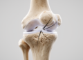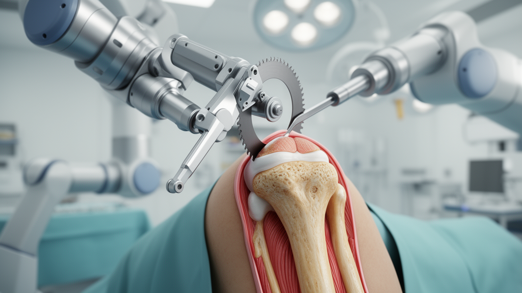
In total knee arthroplasty, achieving precise implant orientation and reconstructing physiological limb alignment are fundamental determinants of joint kinematics, soft tissue equilibrium, and long-term prosthetic durability. In a clinical cohort investigation published in Knee Surgery and Related Research, an orthopedic research team evaluated whether an image-free robotic-assisted surgical platform could enhance bone-cutting precision and alignment accuracy compared to a handheld accelerometer-based navigation unit during ground kinematically aligned total knee arthroplasty. Conducted in individuals presenting with severe varus osteoarthritis, the study provides comparative evidence on how intraoperative robotic guidance refines surgical execution when implementing personalized alignment principles that account for whole-limb load lines extending down to the calcaneus.
Personalized alignment philosophies—including anatomical alignment, unrestricted kinematic alignment, restricted kinematic alignment, and functional alignment—aim to recreate native pre-arthritic articular joint lines, preserve natural constitutional obliquity, and minimize collateral ligament releases. However, systematic meta-analyses have frequently shown that kinematic, anatomical, and functional approaches do not consistently yield definitive clinical superiority over standard mechanical alignment. To resolve these anatomical discrepancies, joint reconstruction specialists introduced the ground mechanical axis, which tracks the continuous weight-bearing line from the center of the femoral head down to the lowest ground-contact point of the calcaneus rather than terminating at the ankle talus center. Because this ground-referenced vector passes directly through the knee center in pristine native limbs, targeting a neutral ground mechanical axis establishes a physiologically stable alignment that accommodates individual hindfoot orientation and natural stance mechanics.
To execute personalized resections with high reproducibility, orthopedic surgeons increasingly incorporate computerized and robotic navigation technologies into their operative workflows. The accelerometer-based portable navigation system evaluated in this investigation—the KneeAlign 2 unit—functions as a compact handheld navigation device providing real-time angular feedback for femoral and tibial osteotomies. Prior literature evaluating this specific robotic platform remained limited primarily to preliminary institutional feasibility reports, leaving open questions regarding its specific precision and clinical utility when executing complex ground kinematically aligned reconstructions.
The clinical trial protocol enrolled thirty-five consecutive adult patients scheduled for ground kinematically aligned total knee arthroplasty using the VELYS robotic-assisted solution between April 2024 and January 2025. One prospective participant withdrew prior to surgery following a confirmed coronavirus infection, resulting in a completed robotic cohort of thirty-four knees. This robotic group was directly compared against a historical control group consisting of thirty-five consecutive patients who underwent identical ground kinematically aligned procedures using the accelerometer-based portable navigation system between May 2020 and June 2023. All procedures were conducted by a single senior surgeon with over fifteen years of arthroplasty experience, utilizing the cruciate-retaining ATTUNE prosthesis. Strict eligibility criteria mandated severe knee osteoarthritis classified as Kellgren-Lawrence grade three or four with a functionally preserved posterior cruciate ligament, confirmed via preoperative epicondylar radiographs and computed tomography, while excluding revision cases, active infections, bilateral surgeries, severe varus exceeding twenty degrees, valgus deformities, flexion contractures exceeding twenty degrees, and preexisting ankle pathologies.
Preoperative surgical planning depended on full-length standing coronal radiographs capturing the entire lower extremity, including the lowest point of the calcaneus under single-leg weight-bearing conditions. Distal femoral bone resection was simulated utilizing the calipered technique, defining the femoral angle between the femoral mechanical axis and a line perpendicular to the distal femoral cut. For proximal tibial planning, surgeons aimed to counterbalance the femoral angle relative to the tibial ground mechanical axis connecting the tibial plateau resection center to the lowest calcaneal contact point. Because intraoperative navigation tools and robotic arrays track the geometric ankle center rather than the laterally positioned calcaneus, surgeons mathematically accounted for this anatomical offset, designated as delta-TA. In a representative surgical plan with a femoral angle of one degree valgus and a calcaneal lateral displacement of two degrees varus, the navigated tibial angle was set to three degrees varus to produce a neutralized post-resection ground mechanical axis while preserving up to ten degrees of natural posterior slope.
All operative procedures were executed using an extension-gap-first workflow following tourniquet inflation to two hundred and fifty millimeters of mercury through a standard medial parapatellar arthrotomy. After verifying functional integrity of the posterior cruciate ligament, the femoral cut was performed with either the robotic solution or the accelerometer-based device using calipered verification, adjusting bone resection depths to match the nine-millimeter implant thickness while compensating for worn cartilage. To quantify intraoperative soft tissue behavior, surgeons employed an offset-type joint tensor secured to the proximal tibia after completing resections. Applying a standardized distraction force of forty pounds, the device measured joint component gaps in millimeters and varus-valgus ligament balance in degrees across flexion angles of zero, ten, thirty, sixty, ninety, and one hundred and twenty degrees under patellofemoral reduction. Medial soft tissue releases of the posteromedial capsule and deep medial collateral ligament were cautiously performed only as needed, prioritizing medial stability alongside a moderately lax lateral compartment and an unconstrained flexion space.
Radiographic assessments performed at one month postoperatively demonstrated superior mechanical accuracy for the robotic cohort across several primary endpoints. Radiographs obtained during single-leg standing revealed that the mean absolute error for the lateral distal femoral angle relative to the surgical plan was 0.7 ± 0.8 degrees in the robotic cohort compared with 1.5 ± 1.1 degrees in the accelerometer navigation cohort, representing a statistically significant enhancement. Similarly, the mean absolute deviation in achieving the neutral ground mechanical axis ratio was significantly smaller in the robotic group at 1.6 ± 1.8 percent versus 3.0 ± 2.4 percent in the control group. Regarding medial proximal tibial angle error, deviations were comparable between groups, recording 0.7 ± 0.7 degrees with the robotic platform and 0.9 ± 0.9 degrees with accelerometer guidance. Crucially, the robotic system completely prevented extreme femoral cutting errors, exhibiting zero outliers exceeding two degrees from the target lateral distal femoral angle, whereas five such outliers occurred in the navigation group. Furthermore, only one robotic patient deviated by more than five percent from the neutral ground mechanical axis target, compared to seven outliers among accelerometer-guided cases.
Intraoperative tensor measurements highlighted subtle yet statistically significant differences in soft tissue dynamics between the two navigation approaches. Across the arc of motion, joint component gaps measured at zero, ten, thirty, sixty, ninety, and one hundred and twenty degrees were 9.1 ± 0.6, 10.9 ± 0.8, 11.3 ± 1.1, 11.1 ± 1.1, 10.5 ± 1.1, and 10.1 ± 0.8 millimeters in the robotic cohort, compared with 9.4 ± 0.8, 11.1 ± 0.8, 11.7 ± 0.9, 11.8 ± 1.1, 11.0 ± 0.8, and 10.6 ± 0.7 millimeters in the accelerometer cohort. Robotic assistance yielded significantly smaller joint gaps during mid-flexion to deep-flexion ranges between thirty and one hundred and twenty degrees. In parallel, coronal ligament balance recordings demonstrated that the robotic group maintained greater lateral laxity throughout knee flexion from ten to one hundred and twenty degrees, recording values of 5.3 ± 1.7 degrees at ten degrees and 3.7 ± 2.0 degrees at deep flexion, compared with 4.3 ± 1.2 degrees and 2.6 ± 2.2 degrees in the navigation cohort. This confirmed that the robotic cut geometry successfully reproduced a balanced envelope featuring medial stability alongside physiological lateral compliance.
Clinical follow-up conducted at one year postoperatively revealed that both surgical cohorts experienced substantial functional recovery, with neither group demonstrating an overall clinical advantage. In the robotic cohort, thirty-two of thirty-four patients completed final clinical testing, as one patient withdrew at three months for malignancy treatment and another passed away at two months from myocardial infarction. Objective assessments revealed no significant differences in postoperative knee extension, which averaged -1.1 ± 2.1 degrees in the robotic group and -2.0 ± 4.0 degrees in the navigation group, or active flexion, averaging 125.9 ± 7.7 degrees versus 126.0 ± 10.1 degrees. Patient-reported metrics evaluated via the 2011 Knee Society Score showed comparable results for objective indicators, satisfaction, and expectations. Although isolated functional activity scores favoured the accelerometer group at 80.2 ± 13.1 points versus 73.3 ± 11.4 points, the net improvement scores from preoperative baseline across all functional subscales and joint motion ranges revealed no statistically significant differences between the two modalities.
Despite the clear mechanical advantages in cutting fidelity and outlier mitigation demonstrated by the robotic system, the authors noted several methodological caveats that restrict generalized clinical interpretations. The non-randomized study design compared a prospective intervention against a historical control group treated during earlier years, introducing potential confounding factors associated with ongoing surgical refinement, progressive institutional familiarity, and subtle evolutions in perioperative management. Furthermore, the overall sample size was relatively small and strictly delimited by statistical power calculations designed around radiological endpoints rather than subtle clinical score variations. The strict exclusion of severe varus, valgus deformities, and hindfoot structural issues limits applicability across broader patient populations. Two-dimensional standing radiographs also cannot capture intricate three-dimensional rotational kinematics, and the reliance on articular surface references may diverge from the true femoral cylindrical axis, underscoring the necessity for extended long-term randomized clinical trials.
Subject of Research: Surgical accuracy and clinical outcomes of robotic-assisted versus accelerometer-based ground kinematically aligned total knee arthroplasty
Article Title: VELYS robotic-assisted solution achieves more accurate bone cutting and neutral ground mechanical axis compared with an accelerometer-based portable navigation system in ground kinematically aligned total knee arthroplasty
Article References: Matsumoto, T., Tsubosaka, M., Nakano, N., Kamenaga, T., Kuroda, Y., Hayashi, S., & Kuroda, R. (2026). VELYS robotic-assisted solution achieves more accurate bone cutting and neutral ground mechanical axis compared with an accelerometer-based portable navigation system in ground kinematically aligned total knee arthroplasty. Knee Surgery & Related Research, 38(1), Article 44. https://doi.org/10.1186/s43019-026-00344-2
Image Credits: AI Generated
DOI: 10.1186/s43019-026-00344-2
Keywords: total knee arthroplasty, VELYS robotic-assisted solution, accelerometer-based portable navigation, ground kinematic alignment, ground mechanical axis, bone cutting accuracy, lateral distal femoral angle, medial proximal tibial angle, varus osteoarthritis, soft tissue balance, joint component gap, ligament laxity, 2011 Knee Society Score, range of motion, implant alignment, cruciate-retaining knee arthroplasty, orthopedic surgery, KneeAlign 2
News Source: Ophelia Keating. (November 18, 2026). Robotic Guidance Improves Bone Cutting Accuracy in Kinematic Knee Arthroplasty. Scienmag.