In an exploratory phase I clinical trial alongside preclinical experiments, researchers investigated non-thermal argon plasma for treating mild-to-moderate atopic dermatitis. Non-thermal argon plasma, a cold physical plasma generated through electrical discharge in argon gas, was examined as a non-pharmacological approach to relieve localized skin inflammation. Conducted at Ajou University Hospital, the investigation evaluated whether brief exposures to this ionized gas could safely alleviate acute eczematous lesions. The trial demonstrated that targeted plasma treatment significantly reduced clinical severity, itch intensity, and life quality impairment compared with a sham device. Parallel laboratory experiments in atopic dermatitis mouse models and cultured human keratinocytes revealed that the physical treatment suppressed inflammatory signaling pathways and diminished oxidative stress within epidermal cells, offering a biologically plausible mechanism for the observed clinical improvements.
Atopic dermatitis is a chronic, relapsing inflammatory skin disorder that affects approximately 2.6 percent of the global population, representing roughly 204 million children and adults worldwide. Findings from the Global Burden of Disease Study 2021 indicate that disease prevalence has risen steadily over the past three decades and will continue growing through 2050, causing considerable nonfatal disability. Although targeted biologic medications targeting interleukin-4 and interleukin-13 have improved outcomes for severe cases, notable therapeutic limitations remain. Biologics require regular injections that provoke anxiety, carry high financial costs, and often permit disease relapses upon discontinuation. Furthermore, because biologic drugs are primarily reserved for uncontrolled moderate-to-severe disease, individuals with mild-to-moderate eczema have few options beyond topical emollients and corticosteroids, highlighting an unmet need for effective, non-invasive therapies.
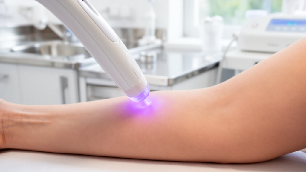
To address this need, researchers evaluated a cold physical plasma device that uses argon as its operating gas. Physical plasma represents an ionized state of matter containing charged particles, electrons, and reactive species generated when electrical energy excites a gas. The device evaluated in the study operated at an input voltage of 15 kilovolts and a frequency of 15 kilohertz, delivering argon at 5 liters per minute. Optical emission spectroscopy across 280 to 920 nanometers confirmed that argon ions dominated the plasma plume. For the clinical trial, investigators designed an inactive sham device that matched the visual exterior, operating sound, and airflow sensation of the active device without generating plasma.
The clinical study was a randomized, double-blind, sham-controlled, parallel-group trial conducted between September 2021 and January 2023 involving fifteen adult patients with mild-to-moderate atopic dermatitis. Mild disease was defined by an Investigator Global Assessment score of 2 and an Eczema Area and Severity Index score between 1.1 and 7.0, whereas moderate disease required an Investigator Global Assessment of 3 and an Eczema Area and Severity Index between 7.1 and 21.0. Participants were randomized two-to-one to receive either active plasma therapy or sham treatment twice weekly for four weeks, totaling nine treatment visits. During each session, five eczematous lesions were irradiated for one minute each. Topical mometasone furoate ointment was permitted only as emergency rescue medication, and patients completed a two-week safety follow-up after concluding the treatment regimen.
Efficacy assessments conducted at week four demonstrated significant clinical improvements in patients treated with active argon plasma compared with the sham cohort. Both an on-site blinded investigator and an independent observer evaluating standardized clinical photographs documented statistically significant reductions in Eczema Area and Severity Index scores among active plasma recipients. Patients in the active group also achieved statistically significant reductions in pruritus on the Numeric Rating Scale and improvements on the Dermatology Life Quality Index. In contrast, the sham group showed no significant changes in these measures. Neither transepidermal water loss nor serum total immunoglobulin E changed significantly after treatment. The authors noted that total immunoglobulin E reflects systemic allergic sensitization that short localized interventions cannot quickly alter, while barrier recovery often lags behind the resolution of inflammation.
To assess whether clinical healing involved microbial shifts, the researchers analyzed skin swabs using 16S ribosomal RNA amplicon sequencing targeting the V3-V4 hypervariable regions. Sequencing on an Illumina MiSeq platform, processed with Cutadapt, DADA2, and QIIME 2 against the SILVA database, showed that plasma altered taxonomic composition. Plasma treatment increased the relative abundance of Lactobacillus and Burkholderia-Caballeronia-Paraburkholderia while reducing Staphylococcus, a genus strongly linked to eczema flares. However, microbial alpha-diversity decreased following treatment, reaching statistical significance only in non-lesional skin, while beta-diversity clustered by treatment group rather than lesion status. Because previous clinical studies commonly associate eczema improvement with increased microbial alpha-diversity, the investigators concluded that these microbiome alterations represented an associated finding rather than the primary causal mechanism driving lesion-specific recovery.
Given the observed reduction in Staphylococcus, the team conducted in vitro experiments using liquid plasma, generated by exposing water or culture media to argon plasma for two minutes per milliliter. When bacterial suspensions were incubated in liquid plasma for ten minutes, Staphylococcus aureus was completely eradicated, whereas Lactobacillus plantarum proved substantially more resistant to plasma-mediated bactericidal activity. Subsequent cell culture assays revealed that co-culturing HaCaT human keratinocytes with Staphylococcus aureus markedly elevated intracellular reactive oxygen species, whereas co-culture with Lactobacillus plantarum produced no such oxidative increase. Furthermore, the results suggested that plasma-induced bacterial shifts might benefit eczema lesions in part by eliminating staphylococcal strains that trigger oxidative stress and reactive oxygen species generation within epidermal keratinocytes.
Because microbiome changes could not fully explain the lesion-specific improvements, researchers used an in vivo mouse model to investigate direct keratinocyte-modulating mechanisms. Eight-week-old male NC/Nga mice had their skin barrier disrupted with 4 percent sodium dodecyl sulfate, followed by application of 100 milligrams of house dust mite cream twice weekly for three weeks to induce atopic dermatitis-like lesions. Mice were divided into untreated controls, house dust mite-treated controls, and house dust mite mice receiving topical argon plasma for two minutes daily over three consecutive days. Histological examination of hematoxylin and eosin-stained skin sections confirmed that argon plasma treatment significantly reduced epidermal thickness and acanthosis. In addition, plasma therapy markedly suppressed dermal inflammatory infiltration, specifically inhibiting the recruitment of allergic effector cells, including mast cells and eosinophils.
Molecular analyses of mouse skin tissues revealed that argon plasma treatment suppressed major allergic signaling cascades. Transcriptomic profiling by RNA sequencing identified significant downregulation of multiple inflammatory cytokines and chemokines, particularly Il33, Ccl2, and Ccl5, with real-time polymerase chain reaction confirming suppressed Il33 transcription. Interleukin-33 functions as an epithelial alarmin that initiates type 2 inflammatory cascades in atopic dermatitis. Western blot analyses demonstrated that plasma application significantly reduced interleukin-33 protein levels, along with downstream signaling intermediates including myeloid differentiation primary response protein 88 and phosphorylated AKT. Immunofluorescence microscopy confirmed a marked reduction in phosphorylated AKT signal intensity within the epidermis of plasma-treated mice. These molecular findings demonstrated that non-thermal argon plasma interrupts the interleukin-33 signaling axis in inflamed epidermal tissue, providing a biological mechanism for reduced cutaneous allergic inflammation.
Because oxidative stress stimulates epithelial interleukin-33 expression, the researchers measured reactive oxygen and nitrogen species in mouse skin. Total reactive oxygen species and reactive nitrogen species, which were significantly elevated in dermatitis-induced mice, were suppressed following argon plasma treatment. Western blot analyses showed that untreated dermatitis skin exhibited elevated baseline levels of nuclear factor erythroid 2-related factor 2 and antioxidant enzymes, including catalase, glutathione peroxidase, superoxide dismutase 1, superoxide dismutase 2, and NAD(P)H:quinone oxidoreductase 1. The authors noted that this endogenous antioxidant response was insufficient to control pathological oxidative levels in untreated dermatitis. Following plasma therapy, antioxidant protein expression declined alongside reduced oxidative species, suggesting that plasma treatment successfully mitigated tissue oxidative stress, thereby reducing the cellular requirement for prolonged compensatory antioxidant enzyme upregulation in resolving skin lesions.
To clarify how plasma modulates antioxidant responses in human skin cells, the researchers conducted in vitro experiments using HaCaT keratinocytes challenged with lipopolysaccharide or hydrogen peroxide. Western blot analyses revealed that while lipopolysaccharide stimulated baseline expression of nuclear factor erythroid 2-related factor 2, liquid plasma treatment further augmented this transcription factor’s expression. Flow cytometry with dihydroethidium demonstrated that liquid plasma significantly reduced intracellular reactive oxygen species in stressed keratinocytes. However, pre-treating keratinocytes with a pharmacological inhibitor of nuclear factor erythroid 2-related factor 2 abolished the antioxidant protection provided by liquid plasma. These in vitro findings supported the concept that argon plasma stimulates the nuclear factor erythroid 2-related factor 2 defense pathway in keratinocytes, thereby boosting antioxidant capacity and counteracting the oxidative stress that drives downstream alarmin expression and eczema pathogenesis.
The authors noted several important study limitations that warrant careful consideration before translating these findings into broad clinical practice. The clinical trial was an exploratory phase I investigation with only fifteen participants, restricting the statistical generalizability of the outcomes. Low bacterial DNA yields from superficial skin swabs precluded high-resolution species-level whole-metagenome sequencing of the microbiome. In addition, the molecular mechanisms involving interleukin-33 and nuclear factor erythroid 2-related factor 2 were established in murine and cell models rather than human skin biopsies in vivo. The trial was also registered retrospectively with the Clinical Research Information Service under registration identifier KCT0008949 due to administrative oversight, though all clinical outcomes were prespecified in the approved protocol. Larger prospective trials with extended follow-up and active drug comparators will be required to confirm long-term clinical efficacy and safety.
Subject of Research: Non-thermal argon plasma therapy for mild-to-moderate atopic dermatitis
Article Title: Non-thermal argon plasma therapy alleviates atopic dermatitis by reducing keratinocyte oxidative stress
Article References: Park, Y. J., Huang, M., Lee, Y. S., Kim, H.-S., Kang, H. Y., & Kim, C.-H. (2026). Non-thermal argon plasma therapy alleviates atopic dermatitis by reducing keratinocyte oxidative stress. Molecular Biomedicine, 7(1), Article 236. https://doi.org/10.1186/s43556-026-00641-y
Image Credits: AI Generated
DOI: 10.1186/s43556-026-00641-y
Keywords: atopic dermatitis, non-thermal argon plasma, cold atmospheric plasma, keratinocytes, oxidative stress, interleukin-33, Eczema Area Severity Index, Dermatology Life Quality Index, Numeric Rating Scale, skin microbiome, Staphylococcus aureus, Lactobacillus plantarum, NRF2 pathway, reactive oxygen species, reactive nitrogen species, NC/Nga mice
News Source: Drew Townsend. (November 18, 2026). Non-Thermal Argon Plasma Eases Mild Atopic Dermatitis in Early Trial. Scienmag.