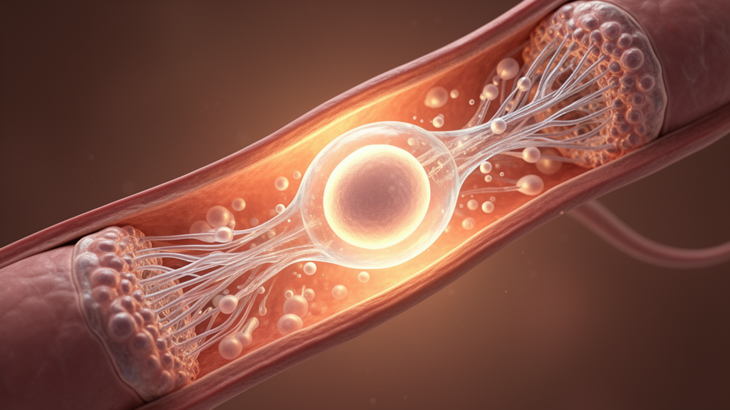
A research team led by investigators at Baylor College of Medicine has captured the key steps of ovulation on video in living mice for the first time. The experimental study, published in Nature Communications, uncovered several previously unrecognized aspects of the ovulatory process and provides new insights into the cellular and tissue dynamics that govern mammalian reproduction. Because abnormalities in ovulation represent the leading cause of female infertility and account for roughly 32 percent of clinical cases, these findings offer potential implications for understanding reproductive disorders.
Ovulation occurs when a mature egg, or oocyte, emerges from an ovarian follicle and travels along the reproductive tract toward the oviduct, where fertilization takes place. Despite widespread recognition of the biological and clinical importance of this transition, observing ovulation directly inside a living animal has historically posed substantial technical challenges. Earlier efforts using endoscopic techniques, laparoscopic imaging, intravital microscopy, or conventional ultrasound were unable to deliver both high spatial resolution and volumetric, three-dimensional video of the event unfolding naturally within intact tissues.
To overcome these observational limitations, the investigators utilized a specialized imaging approach known as intravital optical coherence tomography. Rather than relying on traditional optical microscopy, this computational technology operates on principles conceptually similar to ultrasound but employs light rather than sound waves to acquire large volumes of structural data. The collected signals are computationally processed over several days to reconstruct three-dimensional visualizations of the ovary and surrounding tissues as biological processes occur in real time.
The reconstructed recordings revealed that ovulation does not proceed as an instantaneous rupture, contrary to traditional models that characterized follicle opening as an abrupt event. Instead, the video evidence demonstrated a consistent time lag lasting between 30 seconds and 3 minutes between the initial opening of the ovarian follicle and the eventual release of the oocyte. Following the exit of the egg, the follicular opening closed rapidly within minutes, showing that ovulation is a slow, progressive, and highly coordinated physiological sequence.
In addition to documenting the delayed release, the researchers observed that the typically spherical oocyte undergoes significant mechanical deformation as it passes through the follicular opening. This visible constriction indicates that the egg is physically squeezed during its exit from the follicle. The authors noted that this mechanical deformation may serve as a previously overlooked physical trigger for oocyte activation, as mechanical forces are recognized to influence biochemical signaling, metabolic activity, and gene expression patterns that guide subsequent embryonic development.
The study also resolved longstanding questions regarding how the released oocyte reaches the entrance of the oviduct. Combining intravital imaging with pharmacological testing, the researchers determined that repetitive smooth muscle contractions within the oviduct generate coordinated pulses of periovarian fluid. This fluid movement effectively flushes the newly released oocyte across the periovarian space directly toward the oviduct for potential fertilization.
These observations indicate that smooth muscle dysfunction could lead to oocyte retention in the periovarian space, presenting an uncharacterized mechanism for certain forms of female infertility. Understanding the mechanical and fluid forces required for normal egg transport may ultimately help inform targeted therapeutic interventions for reproductive conditions tied to ovulatory and oviductal motility defects.
The study was led by first author Dr. Kohei Umezu and corresponding author Dr. Irina Larina at Baylor College of Medicine, with additional contributions from researchers at Stevens Institute of Technology and the University of Texas MD Anderson Cancer Center. Funding and support for the research were provided by the Japan Society for the Promotion of Science and the National Institutes of Health.
Subject of Research: Intravital optical coherence tomography imaging of ovulation dynamics in live mice
Article Title: Researchers have captured ovulation on video in live mice
Article References: Researchers have captured ovulation on video in live mice. Original publication (Bibliographic details unavailable.)
Image Credits: AI Generated
DOI: Not provided
Keywords: ovulation, live mice, intravital optical coherence tomography, oocyte release, ovarian follicle, oviduct transport, Baylor College of Medicine, Nature Communications, reproductive biology, smooth muscle contractions, periovarian fluid flow, female infertility, oocyte activation, follicular opening, three-dimensional imaging, computational imaging
News Source: Drew Townsend. (November 17, 2026). High-Resolution Imaging Captures Dynamic Ovulation and Oocyte Transport in Live Mice. Scienmag.