A critical narrative review published in Frontiers in Medicine examines how artificial intelligence and confocal laser endomicroscopy are transforming the diagnosis of obscure small intestine disorders while identifying key structural bottlenecks that continue to impede clinical translation. Investigating deep, tortuous anatomy across several meters of digestive tract has historically challenged gastroenterologists, as conditions such as obscure gastrointestinal bleeding, small bowel tumors, vascular malformations, and early Crohn’s disease present with nonspecific clinical manifestations and are prone to delayed or missed diagnoses. While traditional push enteroscopy reaches only the proximal small intestine leaving substantial blind spots, capsule endoscopy and device-assisted enteroscopy have significantly improved pan-enteric mucosal visualization. However, capsule endoscopy generates tens of thousands of frames per examination that require 30 minutes to two hours of exhausting manual review, whereas device-assisted enteroscopic procedures remain technically demanding and time-consuming.
Artificial intelligence systems trained on deep learning architectures, particularly convolutional neural networks such as YOLO, ResNet, and VGGNet alongside emerging Transformer frameworks, have demonstrated considerable potential in mitigating this reading burden. In capsule endoscopy image recognition, these algorithms operate across distinct operational tiers, successfully automating Tier 1 lesion detection by localizing abnormal mucosal features and partially resolving Tier 2 macroscopic classification by categorizing white-light morphology into erosions, ulcers, polyps, or vascular malformations. Multiple clinical validations indicate that automated reading substantially shortens inspection time and enhances detection sensitivity. For example, in a prospective two-center study enrolling 242 patients evaluated with double-headed capsule endoscopy, automated assistance reduced manual review time from 38.1 minutes to 18.26 minutes, achieving an overall 52 percent reduction in reading duration while preserving a sensitivity of 95.3 percent and delivering a specificity of 96.5 percent compared with 85.3 percent for conventional reading.
Despite these gains in efficiency, a comprehensive appraisal of published data indicates that diagnostic accuracy metrics exhibit considerable variation and asymmetric clinical consequences across different clinical settings. A meta-analysis of fourteen studies reported that algorithm-assisted capsule endoscopy achieved superior sensitivity compared to conventional reading, standing at 92.4 percent versus 86.0 percent, but demonstrated lower specificity, initially reported at 53.7 percent and later corrected to 91.15 percent compared to conventional values of 99.8 percent. Diminished specificity introduces false positives that can trigger unnecessary enteroscopic referrals, escalate hospital costs, and heighten patient anxiety. Furthermore, explainable artificial intelligence techniques such as Grad-CAM saliency maps and attention visualization remain largely absent from published investigations, leaving clinicians without interpretable visual rationales for algorithmic outputs during everyday endoscopic evaluations.
The current body of research on intelligent capsule endoscopy also exhibits notable methodological limitations, specifically regarding lesion spectrum coverage and retrospective database selection bias. Most high-performance algorithms have been trained predominantly on abundant lesions such as ulcers, erosions, and vascular anomalies, leaving clinically critical but less common conditions such as gastrointestinal stromal tumors, lymphangiectasia, and subtle erythema severely underrepresented. Moreover, training datasets frequently undergo aggressive quality screening, systematically excluding frames containing air bubbles, bile staining, food debris, or mucosal visualization below 50 percent. In routine clinical practice, poor visualization affects approximately 13 percent of prepared cases and more than half of unprepared examinations. Because real-world capsule videos routinely contain motion blur and luminal debris, models trained exclusively on sanitized image banks risk optimistic performance overestimation when deployed in authentic clinical environments.
Progress in applying artificial intelligence to device-assisted enteroscopy lags substantially behind capsule endoscopy, primarily due to severe domain shift and dynamic procedural instability. Unlike the relatively stable planar image sequences produced by passive capsule travel, device-assisted enteroscopy involves active water instillation, air insufflation, intestinal wall contact deformation, and rhythmic peristaltic motion that cause pronounced fluctuations in image clarity. Although specialized deep learning systems such as ENDOANGEL-DBE have demonstrated an accuracy of 85 percent in video test sets outperforming endoscopist benchmarks of 77 percent, published investigations remain dominated by single-center feasibility studies. Furthermore, existing enteroscopy models lack cross-platform validation across disparate hardware configurations, including double-balloon, single-balloon, and motorized spiral enteroscopy systems.
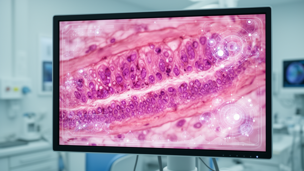
To transcend the boundary of macroscopic mucosal screening and achieve in vivo cellular analysis, researchers have turned to confocal laser endomicroscopy. Utilizing a 488-nanometer blue laser to excite tissue fluorophores and collecting spatial signals reflected from focal planes up to 250 micrometers deep, this technology delivers a spatial resolution of approximately one micrometer, functioning as an optical biopsy. Probe-based confocal systems, which pass miniature imaging probes through standard enteroscope working channels, provide cellular-level visualization of crypt architectures, inter-glandular spaces, microvascular networks, and epithelial barrier function. In clinical cohorts evaluating inflammatory bowel disease, confocal observations of increased epithelial shedding and mucosal fluorescein leakage through defective tight junctions have reliably predicted clinical relapse within twelve months among patients in endoscopic remission.
Nevertheless, small bowel confocal laser endomicroscopy faces a fundamental pharmacological bottleneck that restricts its diagnostic ceiling: fluorescein sodium does not stain cell nuclei. Because intravenous fluorescein contrast primarily highlights the vascular tree and extracellular interstitial space, confocal images reveal microvascular architecture and glandular arrangements but cannot depict nuclear atypia or nuclear-to-cytoplasmic ratios essential for cytological confirmation. While topical nuclear agents like acriflavine have been explored in the upper digestive tract, potential mutagenicity concerns and vast small bowel operating distances impede their mucosal delivery. Consequently, algorithmic feature extraction in small bowel confocal imaging remains confined to glandular morphology and vascular textures, lacking the nuclear descriptors leveraged by digital pathology systems in other anatomical organs.
Beyond pharmacological boundaries, confocal research in the small intestine remains an isolated, underrepresented niche compared with extensive literature accumulated in the esophagus, stomach, and colorectum. Diagnostic parameters used during small bowel examinations are largely borrowed from colorectal frameworks, despite striking anatomical differences between straight colorectal crypt columns and the intricate villus-crypt architecture of the small intestinal mucosa. Current published small bowel confocal cohorts are constrained by single-center designs and modest sample sizes, exemplified by landmark investigations comprising only 20 to 58 patients. Furthermore, physical manipulation within an actively peristalsing small intestinal lumen that is merely one to two millimeters thick frequently causes probe slippage, motion artifacts, or focal contact bleeding, compounding data acquisition scarcity.
To reconcile the structural divide between wide-angle mucosal screening and microscopic tissue characterization, the review authors conceptualize a four-step integrated diagnostic pathway. The framework begins with non-invasive capsule endoscopy to achieve pan-enteric macroscopic visualization, followed by automated deep learning analysis to filter redundant frames, identify subtle lesions, and prioritize urgent pathologies. In the third step, device-assisted enteroscopy navigates directly to algorithmically mapped target coordinates, deploying probe-based confocal imaging to inspect suspected tissues at cellular resolution. Finally, dedicated microscopic neural networks analyze confocal image sequences to quantify goblet cell distribution, inter-glandular distances, and barrier leakage patterns. While this integrated strategy remains a hypothesis-driven conceptual framework awaiting prospective clinical validation, it offers a coherent blueprint for bridging the clinical gap between macroscopic mucosal screening and real-time optical biopsy.
Commercial translation has already begun introducing automated systems into routine practice, marked by regulatory approval in China for OMOM SmartScan, FDA de novo clearance for Navicam ProScan, and 510(k) clearance for Medtronic’s PillCam TOP100 software. In suspected small bowel bleeding, automated reading in a prospective multicenter European cohort increased diagnostic yield from 62.4 percent to 73.7 percent. However, published evaluations remain concentrated almost entirely on technical metrics, such as sensitivity, specificity, and reading times, rather than patient-centered clinical endpoints. Existing studies have not yet determined whether algorithm-assisted capsule endoscopy reduces rebleeding rates, shortens time to targeted intervention, avoids redundant diagnostic procedures, or improves long-term health outcomes.
Confocal laser endomicroscopy has also demonstrated notable prognostic utility in monitoring postoperative Crohn’s disease recurrence. In a prospective pilot investigation of twenty-five patients who underwent ileocolonic resection, probe-based mucosal Watson grades correlated significantly with endoscopic Rutgeerts scores at six to twelve months. Baseline confocal severity scores were significantly elevated among individuals who subsequently experienced endoscopic and clinical relapse over a median follow-up of thirty-eight months. For non-inflammatory conditions, an exploratory single-center study by Dong and colleagues in twenty patients showed that confocal endomicroscopy could clearly differentiate glandular, crypt, and microvascular architectures in small bowel lymphomas, jejunal adenocarcinomas, and Peutz-Jeghers syndrome. Nevertheless, non-inflammatory cohorts remain sparse and underrepresented across the broader endomicroscopy literature.
A critical evidence gap also surrounds whether therapeutic interventions guided by optical biopsy improve ultimate clinical trajectories. While mucosal fluorescein leakage and increased epithelial gap density accurately forecast disease relapse in inflammatory bowel disease, no prospective trials have tested whether escalating or de-escalating medical therapy based on confocal findings alters patient outcomes. Moreover, practical considerations such as formal cost-effectiveness, procedural feasibility in non-academic settings, and broader environmental impacts have yet to be evaluated.
The review authors conclude that the primary bottleneck confronting small bowel artificial intelligence and confocal integration stems not from insurmountable technological feasibility, but from an uneven distribution of academic effort and research resources. Investigating the small intestine entails navigating severe procedural complexity, prolonged examination durations, and higher financial costs for data collection compared to upper gastrointestinal or colorectal endoscopy. Addressing these structural obstacles will require establishing multicenter research networks, unifying acquisition standards, and exploring advanced architectures such as Transformers and self-supervised algorithms that function under data-limited conditions. Finally, the authors emphasize that their narrative review did not perform formal methodological quality grading using QUADAS-2 or AMSTAR-2 tools, and that their proposed four-step pathway remains a conceptual model requiring prospective clinical validation.
Subject of Research: Integration of artificial intelligence and confocal laser endomicroscopy for diagnosing obscure small bowel disorders
Article Title: From macroscopic screening to microscopic characterization: AI and CLE in small bowel disorders
Article References: Zhang, L., & Zhang, W. (2026). From macroscopic screening to microscopic characterization: AI and CLE in small bowel disorders. Frontiers in Medicine, 13, Article 1985297. https://doi.org/10.3389/fmed.2026.1985297
Image Credits: AI Generated
DOI: 10.3389/fmed.2026.1985297
Keywords: small bowel diseases, capsule endoscopy, device-assisted enteroscopy, confocal laser endomicroscopy, artificial intelligence, deep learning, convolutional neural networks, optical biopsy, obscure gastrointestinal bleeding, Crohn's disease, fluorescein sodium, epithelial barrier function, reading efficiency, double-balloon enteroscopy, diagnostic accuracy, biomedical imaging, gastroenterology
News Source: Ophelia Keating. (November 17, 2026). Artificial Intelligence and Confocal Imaging Address Persistent Small Bowel Diagnostic Challenges. Scienmag.