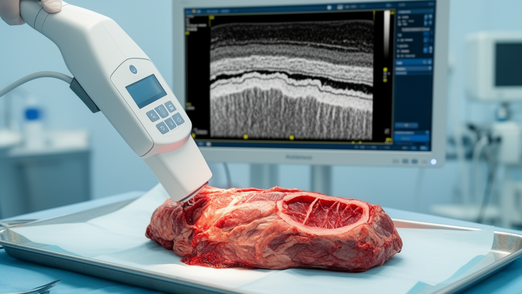
In a clinical feasibility trial assessing optical coherence tomography for margin evaluation following the resection of superficial trunk and extremity soft-tissue sarcomas, an optical imaging technique achieved an overall diagnostic accuracy of 79.8 percent. Investigators at the Ohio State University Comprehensive Cancer Center evaluated twenty excised soft-tissue sarcoma specimens using a handheld optical coherence tomography probe to detect residual neoplastic tissue along surgical boundaries. Across seven trained observers who interpreted standardized test sets, the optical modality demonstrated an overall pooled sensitivity of 83.5 percent and an overall specificity of 76.8 percent against definitive histopathological evaluation. Complete surgical excision with negative margins represents an essential prognostic factor for mitigating local disease recurrence in sarcoma management, yet current standard intraoperative and postoperative diagnostic modalities face considerable sampling and timing limitations. By imaging surgical margins ex vivo immediately after excision, the researchers demonstrated that near-infrared light scattering can distinguish distinct microscopic architectural patterns between healthy tissues and malignant sarcomatous margins.
Although these neoplasms can arise throughout the human body, they appear most frequently within the extremities, where surgical resection serves as the cornerstone of curative intent therapy. When wide resections cannot be achieved, the risk of residual microscopic neoplasia increases, predisposing patients to local recurrence that often necessitates aggressive revision surgeries, reconstructive tissue transfers, or functional limb amputation. Consequently, real-time diagnostic guidance capable of confirming negative margins intraoperatively remains a major objective in orthopedic and surgical oncology practice.
Standard pathological evaluation methods present distinct operational trade-offs during surgical oncology procedures. Definitive histopathological examination of paraffin-embedded tissue remains the diagnostic gold standard, yet processing, sectioning, and staining typically require days to weeks before finalized reports become available to surgical teams. Furthermore, conventional bread-loafing or transverse sectioning trims only evaluate approximately 0.5 to 1.0 percent of the total surgical surface area, potentially missing localized areas of residual disease.
Three patients did not receive optical coherence tomography scanning and were excluded from final evaluations, leaving twenty surgical specimens in the primary analytical cohort. Among these twenty patients, thirteen presented with primary tumors, four presented with recurrent disease, and three underwent resections for metastatic lesions. The study population encompassed ten tumors located in the upper leg, five in the trunk, three in the lower leg, one in the forearm, and one in the upper arm, exhibiting a mean maximum tumor dimension of 8.7 centimeters.
Immediately following surgical excision within the operating theater, the attending surgeon designated the closest surgical margin for optical interrogation. An investigator utilized a commercial 1,300-nanometer spectral-domain optical coherence tomography system equipped with a handheld scanning probe to capture a series of at least twenty cross-sectional optical frames across an identified one-square-centimeter region. The imaged margin was subsequently marked with surgical ink, oriented for pathology, routinely grossed, paraffin-embedded, and sliced into one to five cross sections stained with hematoxylin and eosin. A specialized sarcoma pathologist, strictly blinded to the optical coherence tomography datasets, evaluated the microscopic margin status of the cross sections to establish the ground truth. Overall definitive pathological assessments revealed that fifteen resections achieved negative microscopic margins designated as R0, three cases displayed microscopic positive margins classified as R1, and two resections exhibited gross residual margin involvement classified as R2.
Microscopic architectural analysis revealed distinct optical scattering characteristics across the varied anatomical tissues encountered at surgical boundaries. Normal adipose tissue exhibited a characteristic honeycomb architectural framework characterized by dark, low-scattering intracellular lipid vacuole centers surrounded by brighter, high-scattering cell borders. Skeletal muscle displayed an intermediate to light scattering intensity interspersed with bright, dense linear bands that corresponded histologically to fibrous fascial sheaths enveloping muscle bundles. Intact connective tissue fascia demonstrated the highest scattering density among all normal anatomical structures, appearing nearly brilliant white due to its tightly organized, parallel collagenous bundles. In contrast, sarcomatous tissue displayed high optical scattering intensity that lacked the organized linear hierarchy of healthy fascia and muscle, reflecting the disordered cellular proliferation of malignant tumors, while cellular density concurrently attenuated light penetration, producing reduced imaging depths compared to surrounding normal structures.
To evaluate whether optical criteria could be taught effectively, investigators established a structured visual interpretation curriculum. Optical images and videos derived from the initial five enrolled patients were compared directly against digitized histopathology slides to assemble a standardized training presentation. Seven observers exhibiting varied baseline optical coherence tomography expertise underwent a single thirty-minute virtual instructional session. Following this session, observers evaluated a practice set of ten videos with feedback, followed by a blinded test set comprising twenty-eight videos and two still images sourced from the remaining fifteen surgical patients, including eight vertically mirrored duplicate videos designed to evaluate intraobserver consistency. Observers scored margins using a four-point ordinal confidence scale, where grades of one or two signified tumor absence and grades of three or four denoted positive margins, which were subsequently compiled into two-by-two diagnostic contingency tables.
When pooled across all seven observers, the optical modality yielded an overall correct classification rate of 79.8 percent, correctly categorizing 162 out of 203 margin evaluations with a 95 percent confidence interval ranging from 73.8 to 84.7 percent. The collective sensitivity for detecting malignant tumor cells on the margin reached 83.5 percent, with a 95 percent confidence interval of 74.6 to 89.7 percent, while collective specificity reached 76.8 percent, with a 95 percent confidence interval of 68.2 to 83.6 percent. The overall positive predictive value was determined to be 74.5 percent, and the negative predictive value reached 85.1 percent. Intraobserver reproducibility proved exceptionally strong, yielding perfect kappa agreement values of 1.0 for five of the seven observers, substantial agreement of 0.70 for one proficient observer, and fair agreement of 0.28 for a novice observer, indicating reliable internal consistency across duplicated image sequences.
Despite strong overall performance, substantial variations in diagnostic accuracy emerged across individual observers and tissue conditions. Observer sensitivities ranged from 38.5 percent in a novice interpreter to 100 percent among proficient and advanced observers, while individual specificities spanned 37.5 percent to 100 percent. The researchers observed that neoadjuvant therapies introduced noticeable diagnostic ambiguity; high-density radiation-induced fibrosis exhibited elevated optical scattering and architectural disorganization that closely resembled residual viable sarcoma, making boundary differentiation challenging. Variations in observer turnaround times and intervals following initial instruction also suggested that retention of subtle visual diagnostic criteria may have contributed to differences in classification precision across the testing cohort.
The investigators emphasized several logistical and methodological limitations inherent to this preliminary investigation. The study was restricted to an ex vivo setting examining twenty soft-tissue sarcoma specimens, and each specimen underwent optical coherence tomography scanning at only a single one-square-centimeter marginal location rather than across the entire specimen envelope. Consequently, the study recruited veterinary clinical specialists to complete testing, meaning that diagnostic accuracy among human surgical oncologists operating in active clinical environments remains to be fully verified. Furthermore, the limited one-to-two-millimeter depth penetration of optical coherence tomography restricts assessment to superficial tissue margins, precluding evaluation of deeply seated sub-surface tumor extensions.
Nevertheless, the trial findings demonstrate that optical coherence tomography provides a technically feasible and rapid modality for assessing surgical margin status following soft-tissue sarcoma excision. Whereas standard intraoperative frozen section histological evaluations typically require an average of twenty-six minutes to prepare, section, stain, and interpret, optical coherence tomography image acquisition and interpretation can be completed in five to ten minutes, with experienced investigators scanning targeted margin regions in under sixty-three seconds. The authors concluded that while brief training sessions enable observers to distinguish sarcoma from healthy connective tissue with encouraging diagnostic accuracy, larger prospective clinical studies conducted within active operating theaters are required to evaluate whether intraoperative optical coherence tomography can reliably decrease positive margin rates, prevent secondary revision surgeries, and improve long-term clinical outcomes for sarcoma patients.
Subject of Research: Optical coherence tomography evaluation of soft-tissue sarcoma surgical margins
Article Title: Optical coherence tomography for surgical margin assessment of soft-tissue sarcomas of the extremities and trunk
Article References: Johnson, C., Alexander, J., Mayerson, J., Scharschmidt, T., Grignol, V., Pollock, R., Iwenofu, O. H., Fontes, G. S., Yasenchack, J., Mignucci, J. S., & Selmic, L. E. (2026). Optical coherence tomography for surgical margin assessment of soft-tissue sarcomas of the extremities and trunk. Frontiers in Oncology, 16, Article 1911225. https://doi.org/10.3389/fonc.2026.1911225
Image Credits: AI Generated
DOI: 10.3389/fonc.2026.1911225
Keywords: optical coherence tomography, soft-tissue sarcoma, surgical margins, surgical oncology, histopathology, margin assessment, intraoperative imaging, feasibility trial, biomedical optics, diagnostic accuracy, sensitivity and specificity, frozen section analysis, local recurrence, radiation fibrosis, interobserver variability, resection margins
News Source: Nathaniel Bowman. (November 17, 2026). Optical Coherence Tomography Assesses Soft Tissue Sarcoma Surgical Margins With High Accuracy. Scienmag.