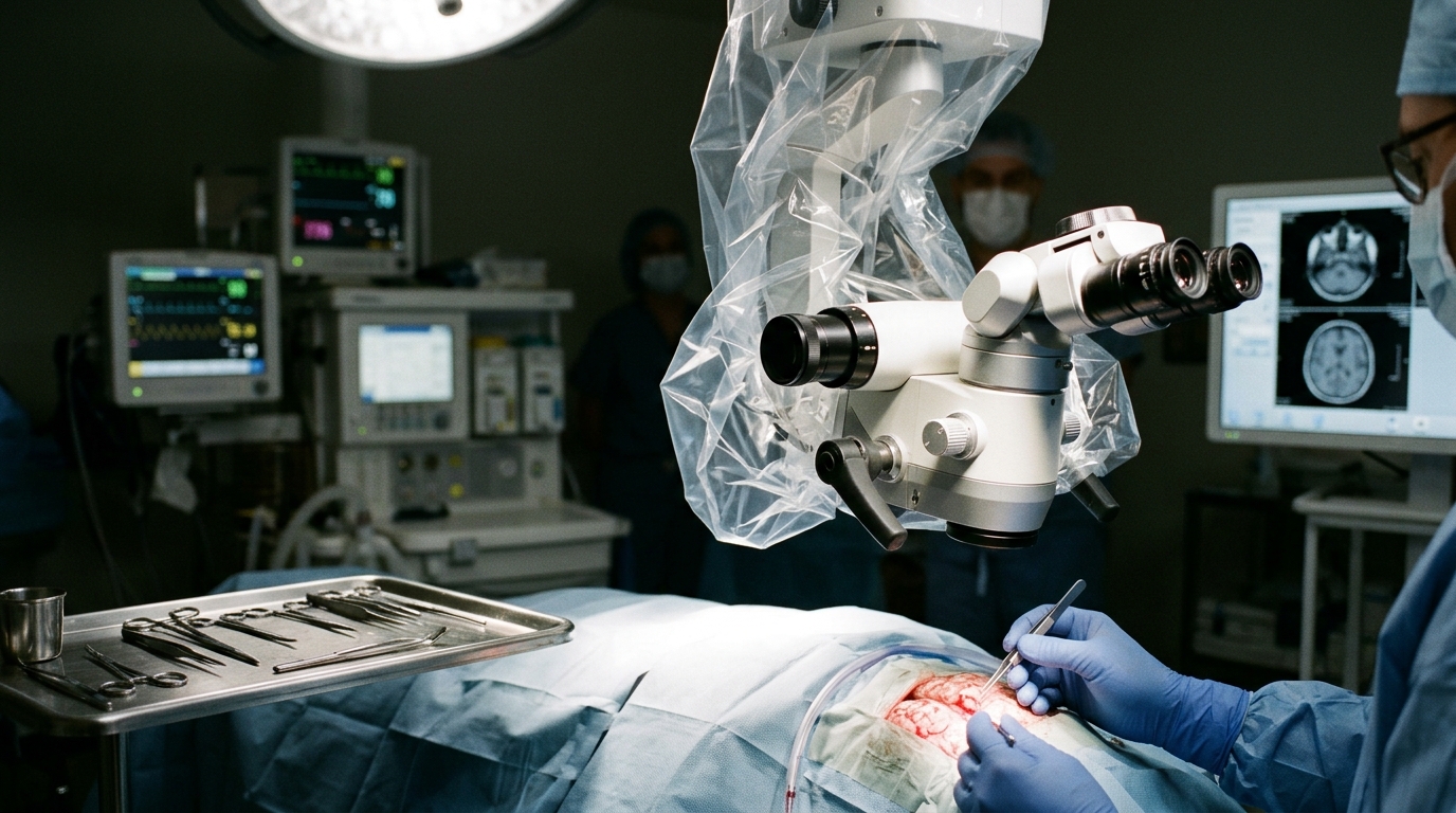
A scoping review evaluating intraoperative detection technologies for diffuse glioma surgery found that while isocitrate dehydrogenase assessment has transitioned toward spatial margin analysis, no validated molecular margin tool currently exists. Researchers examined whether techniques identifying isocitrate dehydrogenase mutation status or its metabolic surrogate, D-2-hydroxyglutarate, have advanced from basic tumour classification to active intraoperative margin guidance. Although surgical extent of resection directly predicts patient survival in isocitrate dehydrogenase-mutant diffuse glioma, infiltrative neoplastic cells routinely extend beyond visually discernible tissue or conventional neuroimaging boundaries, complicating definitive surgical clearance.
To determine whether current analytical systems reliably inform intraoperative surgical decision-making, the investigators implemented a systematic scoping review guided by the PRISMA-ScR framework. The team formulated formal study eligibility around a structured Population, Concept, and Context paradigm. This protocol captured research involving diffuse glioma tissue specimens alongside experimental models, specifically evaluating intraoperatively feasible or proposed analytical platforms configured to measure isocitrate dehydrogenase alterations, D-2-hydroxyglutarate accumulation, or downstream linked diagnostic signals across surgical margin and infiltration zones.
The research team performed systematic electronic literature searches spanning Ovid MEDLINE, Ovid Embase, Web of Science, and Google Scholar, capturing all relevant candidate reports indexed through 15 July 2026. Study selection adhered to rigorous methodological standards, with two independent reviewers completing title, abstract, and full-text screening in duplicate to identify eligible publications. Through this process, the investigators identified and selected thirty-seven unique published studies examining intraoperative identification platforms in diffuse glioma surgery for detailed qualitative synthesis.
Among the thirty-seven included studies, twenty-six investigations specifically focused on isocitrate dehydrogenase-linked detection methodologies. However, only twelve of these twenty-six studies progressed to evaluating molecular or metabolic readouts within a defined spatial context across tissue boundaries. The remaining investigations evaluated bulk tissue samples without resolving boundary-specific margins, illustrating a predominant developmental emphasis on initial diagnostic tumour classification rather than real-time microscopic boundary discrimination during ongoing operative intervention.
Crucially, the review identified substantial diagnostic and analytical evidence gaps across all evaluated margin platforms. None of the included isocitrate dehydrogenase-linked studies established formal diagnostic sensitivity or negative predictive values for local tumour presence at sampled boundary locations verified against an independent, site-matched reference standard. Furthermore, analytical classification thresholds were overwhelmingly developed using bulk tissue or dense tumour core specimens, even though true infiltration edges typically feature markedly reduced cellularity and heterogeneous neoplastic infiltration.
Reported surgical workflows and testing runtimes exhibited extreme variability across different technological modalities. Operational analysis durations spanned from less than three seconds for rapid optical Raman spectroscopy techniques to approximately one hundred minutes for polymerase chain reaction platforms. In addition, sampling strategies and operative margin definitions lacked standardization across institutions. Only two identified studies described extending surgical resection based upon a positive boundary signal, and neither investigation demonstrated measurable, attributable clinical benefit resulting from that additional tissue removal.
The authors concluded that clinical translation of intraoperative isocitrate dehydrogenase detection into validated decision tools demands rigorous, boundary-specific empirical validation. Future translational efforts must establish consistent spatial margin definitions, validate diagnostic thresholds specifically within low-cellularity infiltration zones, and conduct robust site-matched evaluations of both positive and negative analytical readouts. Establishing these rigorous diagnostic benchmarks remains essential before molecular margin readouts can safely guide decisions to resect further tissue or terminate surgical excision.
Subject of Research: Scoping review of intraoperative IDH-linked tumour margin detection in diffuse glioma surgery
Article Title: From tumour classification to tumour margin assessment: a scoping review of intraoperative IDH-linked detection in diffuse glioma surgery
Article References: Sheridan, E. M., Govaerts, C. W., & Price, S. J. (2026). From tumour classification to tumour margin assessment: a scoping review of intraoperative IDH-linked detection in diffuse glioma surgery. Journal of Neuro-Oncology Discovery. https://doi.org/10.1007/s44499-026-00012-9
Image Credits: AI Generated
DOI: 10.1007/s44499-026-00012-9
Keywords: diffuse glioma, IDH mutation, intraoperative detection, tumour margin assessment, extent of resection, D-2-hydroxyglutarate, scoping review, PRISMA-ScR, surgical decision support, Raman spectroscopy, polymerase chain reaction, molecular margins, tumour infiltration, site-matched reference, negative predictive value, low-cellularity tissue, boundary-specific validation
News Source: Nathaniel Bowman. (November 17, 2026). Review Finds Intraoperative IDH Detection Lacks Validated Margin Assessment Evidence. Scienmag.