A refined microsurgical technique utilizing intraluminal polyethylene micro-stents significantly shortens reconstruction time and markedly reduces postoperative morbidity in mouse kidney transplantation, according to a retrospective investigation published in Laboratory Animal Research. Murine renal transplantation represents a foundational laboratory model for investigating ischemia-reperfusion injury, transplant immunomodulation, and the immunological mechanisms driving both acute and chronic graft rejection. However, the procedure remains technically demanding due to the requirement for vascular anastomosis of tiny vessels measuring approximately one millimeter in diameter, paired with the intricate challenge of reconstructing the urinary tract. Because the mouse ureter possesses an exceedingly narrow caliber of only 0.2 to 0.3 millimeters, standard suture-only end-to-end anastomosis is practically unfeasible. Consequently, reconstructive failure historically represents a primary technical barrier that limits experimental success rates, physiological stability, and overall procedural reproducibility.
Historically, two conventional modalities have dominated urinary reconstruction in murine kidney models: the bladder patch technique and the ureter implantation method. The bladder patch technique, which served as the first standardized approach in the field, preserves the recipient’s anatomical anti-reflux mechanism to facilitate long-term observation. Nevertheless, it involves substantial technical complexity and carries high postoperative morbidity, as the donor bladder patch is exceptionally vulnerable to ischemic cuff necrosis. In transplantation models utilizing BALB/c donor strains, necrosis of the bladder cuff and subsequent anastomotic leakage have been documented in up to half of all cases. Conversely, ureter implantation streamlines the reconstructive phase, often allowing completion in under ten minutes, but introduces severe operator dependency. Variation in bladder puncture dimensions, ureteral insertion depth, and anchoring tension frequently results in anastomotic stenosis, graft dislodgement, distal ischemia, or persistent urinary leakage. Furthermore, excessive residual donor ureteral tissue positioned within the bladder lumen can undergo sloughing and necrosis, provoking intractable urinary obstruction and infection.
Beyond operative failure, conventional surgical techniques disrupt the delicate anatomical and physiological architecture of the lower urinary tract. Ureter implantation inherently bypasses the physiological anti-reflux barrier established by the native bladder muscular wall, predisposing recipient animals to vesicoureteral reflux, ascending upper urinary tract infections, and progressive hydronephrosis. While the traditional bladder patch approach retains anti-reflux properties, it does so at the cost of extensive tissue trauma and severe ischemic vulnerability. Similarly, hybrid alternatives like micro-bladder patches carry persistent risks of cuff necrosis without eliminating obstruction, while intraureteral catheterization protocols require parenchymal incisions into the donor renal pelvis that risk iatrogenic tissue damage and late stenosis. To overcome these cumulative technical barriers, the research team designed an end-to-end micro-stenting strategy that bypasses bladder wall manipulation entirely, safeguarding the recipient’s native ureterovesical junction and maintaining the structural integrity necessary for physiological anti-reflux competence.
The study consecutively evaluated orthotopic kidney transplants performed between March 2024 and December 2025 at Shanghai General Hospital under controlled specific-pathogen-free conditions. To rigorously evaluate acute rejection mechanisms under complete major histocompatibility complex mismatching, male BALB/c mice served as organ donors while male C57BL/6 mice weighing between 22 and 25 grams served as recipients across all experimental cohorts. A single microsurgeon performed all operations, eliminating inter-operator variability; notably, while the surgeon possessed substantial microvascular experience from over fifty murine cardiac transplantations, they had no prior experience in rodent urinary tract reconstruction. To prevent chronological skill acquisition from confounding outcome metrics, the three surgical techniques were assigned in a strictly alternating cyclic sequence: intraluminal stenting, bladder patch, and ureter implantation. Of ninety-four consecutive operations, eighty-eight procedures met the predefined criterion of recipient survival past three days post-transplantation following bilateral native nephrectomy, yielding twenty-nine stenting cases, thirty-one bladder patch cases, and twenty-eight ureter implantation cases for comparative analysis.
Standardized microsurgical equipment and protocols were maintained across all procedures under Zeiss stereomicroscopic visualization using specialized micro-forceps, microvascular clamps, and fine nylon sutures. General anesthesia was achieved across donor and recipient cohorts via intraperitoneal administration of ketamine at eighty milligrams per kilogram and xylazine at five milligrams per kilogram. Donor left kidneys were mobilized with intact peri-ureteral mesentery to safeguard local vascularity, perfused in situ via the infrarenal aorta with ice-cold heparinized saline at one hundred units per milliliter until blanching uniformly, and stored in four-degree saline. In recipient animals, bilateral native nephrectomy was conducted, completely removing the left ureter while preserving an eight-millimeter right ureteral stump attached to the bladder junction. End-to-side microvascular anastomoses joined the donor aortic cuff and renal vein to the recipient abdominal aorta and inferior vena cava using 10-0 nylon sutures, restoring graft perfusion prior to initiating urinary tract reconstruction.
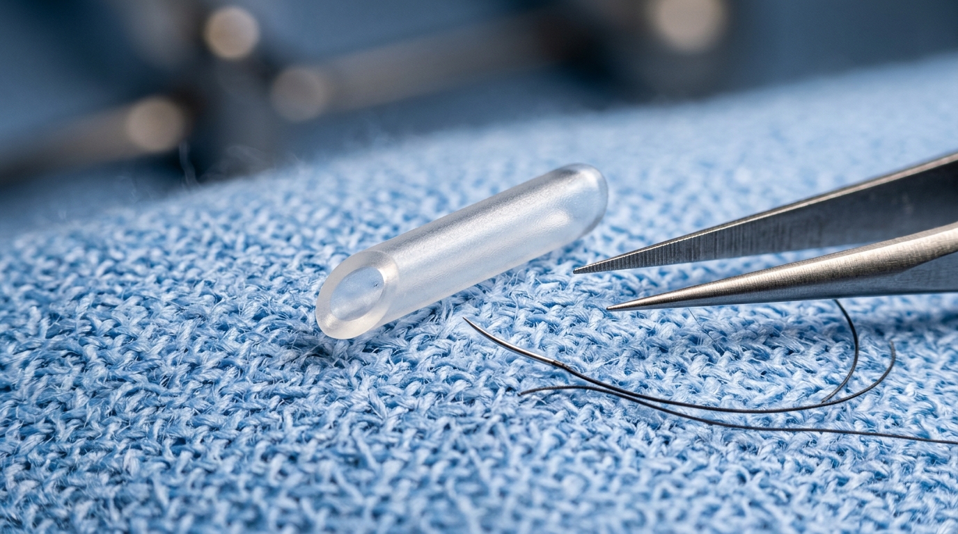
The refined intraluminal stenting protocol relied on custom micro-stents fabricated from medical-grade polyethylene tubing featuring an outer diameter of 0.1 millimeters, an inner diameter of 0.05 millimeters, and a total length of 2.0 millimeters. Both ends of each stent were polished into smooth 45-degree bevels and sterilized with ethylene oxide prior to implantation. During reconstruction, one end of the beveled polyethylene stent was advanced approximately one millimeter into the lumen of the donor ureter and secured externally using a circumferential 10-0 nylon tie. The opposing end of the stent was subsequently docked approximately one millimeter into the proximal lumen of the preserved recipient ureter and anchored with a second 10-0 nylon tie, completing tension-free end-to-end alignment. Finally, the donor and recipient ureteral mesenteries were carefully realigned and approximated with a single interrupted 10-0 suture on each flank, restoring physiological orientation, minimizing anastomotic tension, and preserving vital peri-ureteral microvascular blood flow.
Quantitative assessment of operative duration revealed that intraluminal stenting was markedly faster than established surgical alternatives. Reconstruction time—defined as the interval from graft revascularization to confirmed patency without leakage—averaged 8 minutes 22 seconds (plus or minus 41 seconds) in the stenting group. This duration was significantly shorter than the 33 minutes 42 seconds (plus or minus 3 minutes 37 seconds) required for bladder patch reconstruction and the 11 minutes 11 seconds (plus or minus 1 minute 25 seconds) recorded for ureter implantation. Operative learning curves were evaluated using cumulative sum analysis to pinpoint the inflection point marking surgical proficiency. The intraluminal stenting technique achieved technical plateau after only 10 cases, with mean reconstruction time dropping by 12.3 percent from 9 minutes 5 seconds in the initial phase to 7 minutes 59 seconds in the plateau phase. In contrast, ureter implantation required 14 cases to achieve plateau stability, while the technically laborious bladder patch technique demanded 20 cases, demonstrating that micro-stenting substantially flattens the microsurgical learning curve.
Postoperative fourteen-day survival curves demonstrated clear prognostic advantages for the intraluminal stenting cohort. Mice undergoing intraluminal stenting achieved an overall fourteen-day survival rate of 96.6 percent, with twenty-eight of twenty-nine animals surviving the complete observation window. The sole perioperative loss in this group resulted from delayed vascular anastomotic hemorrhage unrelated to urinary reconstruction, meaning zero urinary-related fatalities occurred in the stented cohort. By contrast, the bladder patch cohort exhibited a significantly lower survival rate of 67.7 percent, involving ten total deaths. One death stemmed from vascular hemorrhage, whereas nine fatalities were directly attributable to urinary tract failure, including four cases of lethal patch necrosis, three instances of fatal peritonitis secondary to anastomotic leakage, and two cases of obstructive renal failure. The ureter implantation cohort demonstrated an intermediate survival rate of 82.1 percent across five total deaths, with four fatalities caused by reconstructive failure consisting of three lethal leakage events and one obstructive ureteral necrosis.
Comprehensive postmortem autopsies and histological examinations conducted on postoperative day fourteen revealed a substantially reduced complication profile following intraluminal micro-stenting. The overall urological complication rate among stented mice was 10.3 percent, representing three isolated events across three animals: one case of mild, self-limiting urinary leakage that resolved spontaneously, and two cases of mild hydronephrosis. Crucially, the stenting cohort experienced zero instances of tissue necrosis, ureteral dislodgement, luminal obstruction, or calculus formation during the fourteen-day evaluation. In comparison, overall complication rates reached 58.1 percent in the bladder patch group and 42.9 percent in the ureter implantation group. The bladder patch cohort accumulated nineteen adverse events across eighteen animals, including eight cases of patch necrosis, five instances of bladder calculi, three leaks, and two cases of hydronephrosis. The ureter implantation cohort suffered twenty-two discrete complications across twelve animals, marked by nine combined necrosis-leakage events, five ureteral dislodgements, four cases of ureteral necrosis, and three severe ureteral obstructions.
Histological evaluation using hematoxylin and eosin staining confirmed that while all three cohorts displayed comparable, severe immune-mediated graft rejection characteristic of full major histocompatibility mismatching, distinct morphological patterns emerged within the renal pelvis. Tissue sections from the ureter implantation group demonstrated marked renal pelvic dilatation, whereas the bladder patch group exhibited mild dilatation. In contrast, grafts reconstructed with intraluminal micro-stents displayed renal pelvic architecture closely resembling normal baseline morphology. This anatomical preservation supports the premise that maintaining an intact recipient ureterovesical junction avoids the retrograde hydrostatic pressure and reflux-induced renal damage frequently observed when the bladder muscularis is breached. By preventing secondary reflux nephropathy and ascending inflammatory confounders, the stenting protocol provides a cleaner immunological baseline for assessing acute rejection phenotypes.
Despite these clear operational benefits, the authors outlined key technical and temporal limitations inherent to the current stenting protocol. A critical limitation centers on stent indwelling duration: in an extended observation animal evaluated beyond the primary experimental endpoint, widespread idiopathic bladder calculi developed at one month post-transplantation. This finding suggests that chronic mechanical irritation from non-degradable polyethylene stents significantly increases urolithiasis risk after four weeks, restricting the current protocol primarily to short-term protocols such as two-week acute rejection assays. To extend the observation window to three or four weeks, the authors proposed testing biodegradable polymer stents engineered to dissolve within two to three weeks, evaluating transurethral stent extraction at postoperative days ten to fourteen, or adjusting systemic hydration regimens. Additional limitations include reliance on a single microsurgeon, exclusive validation in BALB/c and C57BL/6 strains, and the absence of direct urodynamic or functional anti-reflux testing, which remains inferred from anatomical and histological integrity.
Subject of Research: Surgical evaluation of intraluminal micro-stenting for urinary tract reconstruction in murine kidney transplantation
Article Title: Intraluminal micro-stenting improves urinary tract reconstruction in murine kidney transplant model
Article References: Xu, Z.-J., He, L., Tang, Z., Wang, R.-J., Yang, Y., Chen, K.-Y., Ding, Z.-T., Lu, J.-R., Chi, H.-D., Ge, J.-F., & Liu, Z.-H. (2026). Intraluminal micro-stenting improves urinary tract reconstruction in murine kidney transplant model. Laboratory Animal Research, 42(1), Article 39. https://doi.org/10.1186/s42826-026-00301-w
Image Credits: AI Generated
DOI: 10.1186/s42826-026-00301-w
Keywords: Murine kidney transplantation, Intraluminal micro-stenting, Urinary tract reconstruction, Ureteral anastomosis, Polyethylene stents, Microsurgery, Postoperative survival, Hydronephrosis, Urinary leakage, Bladder patch technique, Ureter implantation, Ureterovesical junction, Learning curve, CUSUM analysis, Acute rejection model, BALB/c donors, C57BL/6 recipients
News Source: Drew Townsend. (November 17, 2026). Polyethylene Micro-Stenting Improves Urinary Tract Reconstruction in Mouse Kidney Transplants. Scienmag.