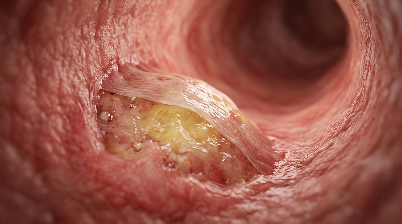
In an exploratory pilot investigation examining high-risk cardiac presentations, researchers evaluating thirty individuals experiencing acute ST-elevation myocardial infarction identified that microstructural vulnerability within culprit coronary plaques was descriptively linked to a higher frequency of short-term adverse cardiovascular events following emergency percutaneous revascularization. The investigation, conducted at a single tertiary care facility and reported in the Journal of Cardiovascular Imaging, specifically explored how intravascular optical coherence tomography assessments of plaque architecture behave alongside macroscopic calculations of residual disease burden quantified through the residual SYNTAX score. Across the evaluated clinical cohort, individuals presenting with optical coherence tomography-defined thin-cap fibroatheroma accounted for seven out of nine recorded major adverse cardiac events documented during a three-month post-procedural surveillance window, regardless of whether angiographic post-intervention scoring indicated reasonably complete or incomplete coronary revascularization.
Acute ST-elevation myocardial infarction persists as a primary contributor to global cardiovascular morbidity and mortality, characterized pathophysiologically by the acute rupture of vulnerable atherosclerotic plaques, subsequent occlusive thrombus development, and severe ischemic injury to surrounding myocardial tissue. While emergency percutaneous coronary intervention represents the established clinical standard for restoring coronary perfusion, surviving patients remain subject to secondary clinical vulnerability governed simultaneously by untreated macroscopic obstructive coronary lesions and intrinsic biological instability within the vessel wall. Interventional cardiology routinely employs the residual SYNTAX score on post-procedure completion angiograms to quantify untreated macroscopic stenosis across remaining coronary segments. Historically, residual score values equal to or less than four serve as an established benchmark denoting reasonable revascularization with low long-term adverse ischemic risk, while higher scores correlate with elevated rates of recurrent myocardial infarction, heart failure hospitalizations, and overall mortality.
Despite the utility of macroscopic angiographic scoring, standard angiography remains incapable of characterizing microscopic plaque composition or biological fragility. To evaluate microstructural vascular architecture in vivo, investigators deployed high-resolution intravascular optical coherence tomography, an imaging modality that utilizes automated catheter pullbacks alongside continuous contrast displacement of luminal blood to yield cross-sectional vascular renderings with an axial resolution spanning ten to twenty micrometers. This imaging fidelity enables the direct identification and measurement of thin-cap fibroatheroma, a high-risk morphological structure characterized by an extensive lipid core and an extremely attenuated fibrous cap. Prior prospective imaging studies have linked thin-cap fibroatheroma and lipid pools located near stent margins to impaired vascular healing, incomplete strut endothelialization, and elevated clinical vulnerability, establishing a biological rationale for combining microstructural assessments with macroscopic luminal evaluation.
The retrospective single-center pilot protocol was conducted between April 2023 and March 2024 following formal ethical approval from the Institutional Ethics Committee of Nizam’s Institute of Medical Sciences. A total of 142 individuals presenting with acute ST-elevation myocardial infarction were consecutively screened against rigorous diagnostic criteria, requiring persistent chest discomfort, biomarker elevation, and diagnostic electrocardiographic changes such as ST-segment elevation exceeding 0.1 millivolts across contiguous leads or new-onset left bundle branch blocks. Eligible participants underwent culprit vessel optical coherence tomography utilizing the C7-XR and ILUMIEN platform paired with Dragonfly catheter equipment. To isolate straightforward procedural mechanics within an initial descriptive cohort, the investigative team excluded individuals presenting with cardiogenic shock, profound renal impairment, left main coronary trunk lesions, chronic total occlusions, extreme vessel tortuosity, and severe radiopaque contrast allergies, resulting in a finalized exploratory cohort of thirty patients.
Within the finalized study population, offline optical coherence tomography recordings were scrutinized independently by two cardiologists blinded to subsequent patient outcomes. In accordance with established international consensus definitions, thin-cap fibroatheroma was defined as a lipid-rich atherosclerotic plaque demonstrating a maximal lipid arc exceeding ninety degrees and a thinnest fibrous cap measurement beneath sixty-five micrometers. Concurrently, post-intervention completion angiograms were reviewed to calculate the residual SYNTAX score using the established cutpoint of four. By cross-referencing microstructural plaque morphology against macrovascular revascularization status, the researchers stratified the cohort into four distinct clinical groups: group zero comprised seven patients lacking thin-cap fibroatheroma who had residual scores greater than four; group one comprised seven patients lacking thin-cap fibroatheroma with residual scores of four or lower; group two comprised eleven patients presenting with confirmed thin-cap fibroatheroma and residual scores greater than four; and group three comprised five patients presenting with thin-cap fibroatheroma alongside residual scores of four or lower.
Baseline cohort demographics demonstrated a mean age of 54.5 ± 10.1 years, a marked male predominance of 83.3 percent representing twenty-five individuals, and an anterior wall infarction presentation in 46.7 percent of the cohort. Cardiovascular comorbidity was highly prevalent, with hypertension documented in 53.3 percent, diabetes mellitus in 40.0 percent, dyslipidemia in 36.7 percent, and active tobacco smoking in 33.3 percent of participants, while baseline mean left ventricular ejection fraction stood at 46.5 ± 8.3 percent. Culprit lesion optical coherence tomography identified thin-cap fibroatheroma in sixteen of the thirty individuals, reflecting an overall prevalence of 53.3 percent. Direct caliper measurements confirmed significant microstructural disparities between groups, with affected patients exhibiting marked fibrous cap thinning measuring 55.0 ± 2.2 micrometers in group two and 54.4 ± 5.4 micrometers in group three, compared to 89.6 ± 10.8 micrometers in group zero and 90.8 ± 13.2 micrometers in group one.
The primary clinical endpoint was major adverse cardiac events assessed over a three-month post-procedural surveillance window, defined as a composite outcome encompassing all-cause death, recurrent myocardial infarction, ischemic stroke, target lesion revascularization, and hospitalization for heart failure. Across the entire thirty-patient cohort, nine individuals experienced primary clinical endpoints, establishing an overall cumulative adverse event incidence of 30.0 percent. The specific event breakdown across three months documented heart failure hospitalization in three patients representing 10.0 percent, ischemic stroke in two patients representing 6.7 percent, target lesion revascularization in two patients representing 6.7 percent, recurrent myocardial infarction in one patient representing 3.3 percent, and all-cause mortality in one patient representing 3.3 percent, with all clinical occurrences verified by independent adjudicating cardiologists.
Evaluating clinical outcomes across the discrete anatomical and biological strata revealed notable descriptive variations across the four patient groups. Among patients without culprit thin-cap fibroatheroma, event rates remained stable regardless of residual coronary stenosis, with major adverse cardiac events occurring in one of seven patients representing 14.3 percent in group zero and one of seven patients representing 14.3 percent in group one. Conversely, higher event concentrations appeared among individuals harboring vulnerable plaque architecture: events were documented in four of eleven patients representing 36.4 percent in group two and three of five patients representing 60.0 percent in group three. When analyzing biological plaque characteristics in isolation, patients possessing thin-cap fibroatheroma experienced an aggregate event frequency of 43.8 percent representing seven of sixteen individuals, compared to 14.3 percent representing two of fourteen individuals among those without vulnerable plaques, accounting for 77.8 percent of all recorded adverse clinical outcomes.
Despite the apparent numerical concentration of events among patients presenting with vulnerable plaque features, statistical comparisons across the four strata did not achieve formal significance. Categorical evaluation utilizing the Fisher exact test yielded a probability value of P = 0.23, reflecting sparse cell frequencies across the small cohort. Furthermore, Kaplan–Meier event-free survival curves depicting clinical trajectories over ninety days demonstrated no statistically significant separation between the four groups under Mantel-Cox log-rank evaluation, generating a chi-square value of 3.08 and a corresponding P-value of 0.38. Because the overall cohort was limited to thirty patients with only nine cumulative events, multivariable Cox proportional hazards regression and formal predictive risk modeling were deliberately omitted to prevent severe mathematical overfitting, maintaining the observations as strictly exploratory and hypothesis-generating.
The authors contextualized the apparent paradox observed within group three, where patients achieving reasonable macrovascular revascularization with residual scores of four or lower demonstrated a descriptive event rate of 60.0 percent compared to 36.4 percent in group two. Interventional cardiologists emphasized that with an overall denominator of merely five patients, individual clinical occurrences exert a disproportionate mathematical influence, shifting the group event frequency by twenty percentage points per occurrence. Furthermore, two of the three recorded events in group three consisted of repeat target lesion revascularizations, indicating that the disparity reflects extreme sampling variability within an underpowered subset rather than demonstrable biological resistance to coronary revascularization. This pattern reinforces the fundamental limitation that observational pilot comparisons cannot support causal inferences regarding treatment response.
Crucially, the study authors highlighted key methodological constraints and substantial selection bias that fundamentally shape data interpretation. The observed 30.0 percent cumulative event rate is exceptionally high for a comparatively young cohort with a mean age under fifty-five years, and the incidence of ischemic stroke exceeded recurrent myocardial infarctions, an outcome distribution distinctly atypical for unselected acute coronary syndrome registries. This pattern underscores that optical coherence tomography was not applied systematically across all presenting admissions but was selectively utilized in complex cases. Furthermore, intravascular imaging was restricted exclusively to acute culprit lesions, leaving nonculprit vessel biological vulnerability unmeasured despite known systemic risks. The authors concluded that while their exploratory observations support the biological rationale for integrating intravascular plaque characterization with macroscopic residual scoring, large prospective multicenter registries are essential to determine whether dual anatomical-biological risk stratification provides independent prognostic utility in routine post-infarction care.
Subject of Research: Integration of optical coherence tomography plaque assessment and residual SYNTAX score in ST-elevation myocardial infarction
Article Title: Residual SYNTAX score and optical coherence tomography–derived thin-cap fibroatheroma in patients with ST-elevation myocardial infarction undergoing percutaneous coronary intervention: an exploratory descriptive pilot study
Article References: Syed, M., Karuru, U. D., Arumulla, S., Doddi, N., Rongali, P., & Nuthalapati, R. K. (2026). Residual SYNTAX score and optical coherence tomography–derived thin-cap fibroatheroma in patients with ST-elevation myocardial infarction undergoing percutaneous coronary intervention: an exploratory descriptive pilot study. Journal of Cardiovascular Imaging, 34(1), Article 42. https://doi.org/10.1186/s44348-026-00104-6
Image Credits: AI Generated
DOI: 10.1186/s44348-026-00104-6
Keywords: ST-elevation myocardial infarction, optical coherence tomography, thin-cap fibroatheroma, residual SYNTAX score, percutaneous coronary intervention, major adverse cardiac events, plaque vulnerability, culprit lesion, intravascular imaging, fibrous cap thickness, revascularization, coronary artery disease, cardiovascular imaging, interventional cardiology, pilot study, target lesion revascularization
News Source: Ophelia Keating. (November 17, 2026). Coronary Plaque Vulnerability Descriptively Linked to Adverse Events After Heart Attack. Scienmag.