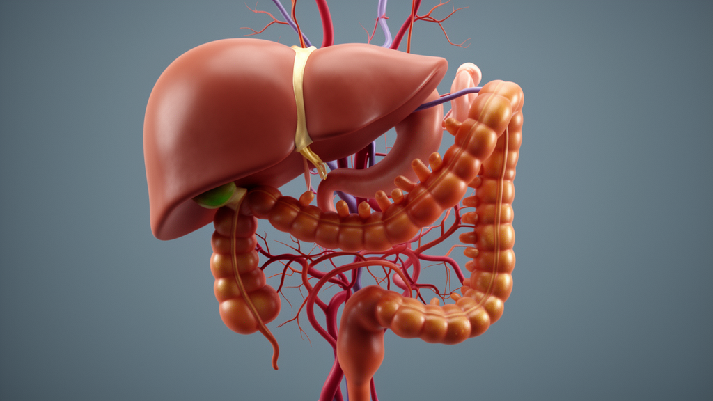
A case report published in Frontiers in Nutrition describes a rare and severe hepatic presentation in a 38-year-old woman with celiac disease. The patient experienced conjugated jaundice and steatohepatitis after approximately 18 years of nonadherence to a gluten-free diet. The authors note that while mild aminotransferase elevation is a recognized manifestation of celiac disease, conjugated jaundice accompanied by steatohepatitis is uncommon. This report illustrates a rare hepatic phenotype associated with prolonged gluten exposure and severe malnutrition, highlighting the potential for reversible liver injury in the context of chronic dietary lapse.
The patient, a Tunisian woman diagnosed with celiac disease at age two, reported complete discontinuation of the gluten-free diet for nearly two decades. Four months before admission, she developed progressively worsening diarrhea and an unintentional 20-kg weight loss. Two weeks prior to presentation, she noticed jaundice and dark urine without pruritus. On examination, her body mass index was 16 kg/m², indicating severe underweight status. She denied alcohol consumption, medication use, or exposure to recreational drugs or environmental toxins. The clinical timeline revealed a clear progression from gastrointestinal symptoms to overt hepatic dysfunction, culminating in admission with significant nutritional compromise.
Initial laboratory testing revealed predominantly conjugated hyperbilirubinemia, with total bilirubin at 192 μmol/L and conjugated bilirubin at 168 μmol/L. Liver enzymes were elevated, with aspartate aminotransferase at 2.3 times the upper limit of normal, alanine aminotransferase at 1.8 times, alkaline phosphatase at 1.3 times, and gamma-glutamyl transferase at 3.3 times the upper limit of normal. Additional findings included anemia, hypoalbuminemia, hyponatremia, and hypokalemia, which supported active malabsorption and severe nutritional compromise. Serologic testing for hepatitis A, B, C, and E was negative, and prothrombin activity was 78%. These quantitative markers established the severity of the hepatic and nutritional derangement at the time of admission.
Diagnostic imaging and histology provided critical insights into the liver pathology. Abdominal ultrasonography showed a steatotic liver with patent hepatic vessels and no bile-duct dilation, effectively ruling out biliary obstruction. Duodenal biopsies demonstrated villous atrophy, confirming active enteropathy. Liver biopsy revealed macrovesicular steatosis involving approximately 40% of hepatocytes, neutrophilic inflammation, focal hepatocyte necrosis, and moderate fibrosis. No cholangitis was identified. The integrated diagnosis was gluten-responsive, celiac-associated liver injury with malnutrition-related steatosis and cholestatic jaundice. The authors noted that classic isolated celiac hepatitis alone was insufficient to explain the marked conjugated hyperbilirubinemia and steatosis observed in this patient.
Therapeutic intervention focused on strict gluten withdrawal and nutritional rehabilitation. A strict gluten-free diet was resumed immediately, accompanied by correction of electrolyte abnormalities and nutritional deficits. Due to severe malnutrition and inadequate early intake, parenteral nutrition was administered for seven days during the third week of hospitalization. The intravenous lipid component was a 100% soybean-oil emulsion. Corticosteroids and other immunosuppressive agents were not initiated because the autoimmune hepatitis workup and histology were not supportive. This conservative management approach aimed to address the underlying dietary nonadherence and nutritional stress without introducing additional pharmacological variables.
During and shortly after the parenteral nutrition course, aminotransferases rose transiently, peaking at 8.2 times the upper limit of normal for AST and 5.6 times for ALT on day 33. An adapted RUCAM score of 4 indicated only a possible contribution of the parenteral nutrition to this secondary flare. The authors emphasized that RUCAM was developed for drug exposures rather than multicomponent parenteral nutrition, so the result was interpreted as supportive but not definitive. Bilirubin levels had already begun to decline around the end of the parenteral nutrition course, peaking at 215 μmol/L on day 19 and decreasing to 49 μmol/L by day 33. This divergence between improving bilirubin and the transient enzyme flare argued against attributing the entire hepatic syndrome to parenteral nutrition.
Follow-up outcomes demonstrated significant clinical and biochemical recovery. By two months, diarrhea and jaundice had resolved, and the patient had gained weight with progressive improvement in general condition. At nine months, her body mass index had increased to 25 kg/m². AST, ALT, total bilirubin, and alkaline phosphatase levels returned to normal ranges. Gamma-glutamyl transferase remained minimally elevated at 1.5 times the upper limit of normal. Adherence at follow-up was assessed from the patient’s report and the clinical course, though it was not objectively confirmed by serial serologic measurements. The sustained response to gluten withdrawal and nutritional restoration supported the final diagnosis of a reversible hepatic phenotype.
The authors discuss the pathophysiologic interpretation, proposing a multifactorial mechanism involving active enteropathy, barrier dysfunction, and dysbiosis. These factors may increase portal delivery of microbial products and pro-inflammatory cytokines, leading to hepatic inflammation. Severe malabsorption and rapid weight loss likely acted as additional metabolic stressors, impairing hepatic lipid handling and favoring macrovesicular steatosis. The authors note that while parenteral nutrition can contribute to steatosis, this patient had established cholestatic steatohepatitis before its administration. The short duration of parenteral nutrition and the simultaneous fall in bilirubin further support the view that it did not cause the primary hepatic syndrome.
Limitations of the case report include the absence of quantitative celiac serology at admission and during follow-up, which precluded serologic confirmation of disease activity and dietary adherence. Additionally, magnetic resonance cholangiopancreatography was not documented, limiting the definitive exclusion of structural large-duct disease such as primary sclerosing cholangitis. Serum ceruloplasmin, alpha-1 antitrypsin, and iron overload markers were unavailable, meaning these inherited or metabolic conditions could not be biochemically excluded. The authors acknowledge that the diagnosis rests on clinicopathologic integration and a clinical dechallenge response rather than on a disease-specific test. These limitations temper the strength of the causal attribution to gluten nonadherence.
This case broadens the recognized hepatic spectrum of celiac disease, which typically presents with mild hepatocellular enzyme elevation. The presence of clinically evident conjugated jaundice, substantial steatosis, and fibrosis in this patient highlights the potential for severe liver dysfunction in the setting of prolonged dietary nonadherence. The authors emphasize the importance of considering active celiac disease in patients with unexplained cholestatic liver abnormalities and steatosis, particularly when accompanied by diarrhea, weight loss, and nutritional deficiencies. Conversely, lack of improvement after documented gluten-free diet adherence should prompt renewed evaluation for other liver diseases. This report underscores the need for careful monitoring and comprehensive diagnostic workup in celiac patients with atypical hepatic presentations.
To evaluate other potential drivers of acute deterioration, clinicians screened for occult malignancies and autoimmune liver conditions. Because long-standing uncontrolled celiac disease carries a risk of lymphoproliferative complications, the team assessed serum markers and performed imaging. Serum lactate dehydrogenase and beta-2 microglobulin were normal, while thoracoabdominopelvic computed tomography revealed no abnormal mass lesions or adenopathy. Bone-marrow examination showed no evidence of malignancy, and mucosal biopsies from upper endoscopy and ileocolonoscopy excluded lymphoma. Serologic testing demonstrated antinuclear antibodies at 1:400 with a speckled pattern, but assays for anti-smooth muscle, antimitochondrial M2, anti-sp100, anti-gp210, liver-kidney microsomal type 1, soluble liver antigen, and liver cytosol type 1 were negative. With normal immunoglobulin G concentrations and normal serum globulins, the simplified International Autoimmune Hepatitis Group score was 4, falling below the threshold for probable autoimmune hepatitis.
The authors contextualized these findings within broader hepatology literature examining hepatic involvement in celiac disease. Biopsy data from a cohort of 36 patients documented hepatic steatosis in 50 percent, steatohepatitis in 19 percent, and non-specific lobular or portal inflammation in 25 percent, while an underlying coexisting hepatic condition was identified in 75 percent. Earlier single-case publications by Schmidt, Cassagnou, Kaukinen, and Gaur similarly reported reversible liver injury, severe hepatic impairment, or marked regression of steatohepatitis and fibrosis following strict dietary intervention. Furthermore, the report distinguished this presentation from typical metabolic dysfunction-associated steatotic liver disease. Although a 2025 cross-sectional study observed hepatic steatosis in 34 percent of adults with celiac disease and identified higher body mass index as the primary predictor, the present patient developed severe steatohepatitis amidst pronounced wasting and catabolic weight loss. This difference underscores that celiac-associated fat accumulation cannot simply be attributed to conventional cardiometabolic risks.
Socio-cultural and dietary dynamics played an important role in the clinical trajectory. In Mediterranean communities, wheat-based products form an integral part of everyday meals and social customs, while certified gluten-free foods remain costly and inconsistently accessible. In her personal account, the patient explained that she originally abandoned dietary restrictions during adolescence because adherence felt socially isolating and burdensome within a wheat-centric household. Crucially, she experienced an extended asymptomatic period spanning several years after stopping the diet, which reinforced the false impression that dietary compliance was unnecessary. Following her severe illness, successful nutritional recovery and the complete disappearance of chronic diarrhea and jaundice convinced her to adhere strictly to gluten avoidance over the long term.
Additional analytical nuances defined the diagnostic course and longitudinal monitoring. During the day-33 enzyme peak, alkaline phosphatase reached approximately 1.6 times the upper limit of normal alongside aminotransferase elevations, producing an R ratio of approximately 3.5 that signified a mixed pattern of liver injury rather than purely hepatocellular or cholestatic damage. Total bilirubin dropped steadily from 49 μmol/L on day 33 to 28 μmol/L by day 66. Pathologists faced specific documentation limits, as the retrievable medical records lacked the precise number and anatomical distribution of duodenal specimens between the bulb and distal duodenum, and did not record a formalized Marsh-Oberhuber histologic grade.
Subject of Research: Reversible cholestatic steatohepatitis associated with prolonged gluten-free diet nonadherence in celiac disease
Article Title: Case Report: Reversible cholestatic steatohepatitis after two decades of gluten-free diet non-adherence in celiac disease
Article References: Sallem, O. K., Bouhlel, W., Ben Chaabene, N., & Safer, L. (2026). Case Report: Reversible cholestatic steatohepatitis after two decades of gluten-free diet non-adherence in celiac disease. Frontiers in Nutrition, 13, Article 1942195. https://doi.org/10.3389/fnut.2026.1942195
Image Credits: AI Generated
DOI: 10.3389/fnut.2026.1942195
Keywords: celiac disease, cholestatic steatohepatitis, gluten-free diet, dietary nonadherence, hepatic manifestations, malnutrition, conjugated jaundice, liver biopsy, parenteral nutrition, clinical case report, gut-liver axis, villous atrophy, nutritional rehabilitation, Tunisia, hepatobiliary disorders
News Source: Ophelia Keating. (November 14, 2026). Celiac Disease Linked to Reversible Liver Damage in Case Report. Scienmag.