In a peer-reviewed research study published on October 8, 2026, in volume 18 of the journal Aging, investigators demonstrated that small extracellular vesicles released by senescent human cells transmit signals that induce cellular senescence in neighboring healthy cells. The research, titled “Small extracellular vesicles as mediators of paracrine senescence in cellular models of aging,” was conducted by equal-contributing first authors Aina M. Llabrés-Mas, Antonio Merino-Navarro, and Marta Menéndez-García alongside corresponding author Ana O’Loghlen, all affiliated with the Spanish National Research Council, known as CSIC, in Madrid, Spain. Utilizing controlled human cellular models of aging, the scientific team established that these membrane-enclosed nanoparticles can trigger stable cell-cycle arrest and activate DNA damage responses in recipient cells without stimulating the classical inflammatory signaling pathways conventionally linked to cellular aging. These experimental observations uncover an alternative biological pathway through which non-proliferating aged cells communicate with their surroundings in laboratory culture systems.
Cellular senescence describes a biological process wherein cells enter an enduring state of permanent growth arrest following exposure to physiological stress, structural damage, or specific developmental cues. Under physiological conditions, this growth arrest serves an essential protective function by halting the proliferation of damaged cells and facilitating localized tissue repair mechanisms. Nevertheless, the accumulation of senescent cells over extended periods is closely tied to systemic aging, chronic inflammation, and a wide array of age-associated pathologies. Historically, researchers have understood that senescent cells alter surrounding tissue environments through the senescence-associated secretory phenotype, commonly abbreviated as SASP. This secretory program releases a complex mixture of soluble signaling molecules, including pro-inflammatory cytokines, chemokines, and growth factors, that can trigger paracrine senescence by coercing adjacent, previously dividing cells into growth arrest. However, this classical inflammatory pathway is not universal across all forms of senescence, creating an important scientific puzzle regarding how non-inflammatory senescent cells influence surrounding tissue compartments.
To investigate this question, the CSIC research team focused on cells expressing elevated amounts of p16INK4A, a key protein responsible for regulating normal cell-cycle progression. Cells that enter senescence driven by p16INK4A maintain a stable growth-arrested state while producing negligible levels of standard inflammatory SASP factors. For their experimental framework, the researchers modified human primary IMR-90 fibroblasts to ectopically express p16INK4A. They systematically compared these modified cells against actively proliferating control fibroblasts carrying an empty vector and cells expressing oncogenic H-RASG12V, which served as a model for senescence accompanied by an inflammatory SASP. Quantitative real-time polymerase chain reaction, or qPCR, confirmed marked elevations of CDKN2A mRNA levels in the p16-transduced fibroblasts across three independent experiments. Furthermore, expression heatmaps of transcripts related to SASP confirmed that while RAS-transduced fibroblasts robustly upregulated inflammatory markers, p16INK4A-expressing fibroblasts exhibited minimal transcriptional activity for these genes. Enzyme-linked immunosorbent assays, or ELISA, measuring conditioned culture media across three experiments with mean and standard error of the mean confirmed that RAS-expressing cells secreted substantial amounts of interleukin-6 and interleukin-1beta, whereas p16-expressing fibroblasts released virtually undetectable amounts.
Despite displaying no classical inflammatory response, the p16INK4A-expressing fibroblasts released significantly greater quantities of small extracellular vesicles into their culture medium than proliferating control cells. Small extracellular vesicles, frequently designated as sEVs, represent membrane-bound particles that transport biologically active lipids, proteins, and nucleic acids between cells, functioning as biological messengers. The researchers harvested and purified these vesicles from conditioned media using ultracentrifugation. To characterize the isolated vesicular populations, the team performed Nanoparticle Tracking Analysis, commonly referred to as NTA, which verified that p16-expressing fibroblasts produced markedly higher particle counts than control cells. Concurrently, NTA size profiling and parametric Student’s t-tests revealed that average particle diameter showed no significant differences between control, RAS, and p16 groups. Immunoblotting performed across three biological replicates, loading 3×10^9 vesicles per lane, confirmed the presence of established vesicle markers, including ALIX, TSG101, and CD9. Complementary flow cytometry utilizing aldehyde-sulphate latex beads demonstrated surface enrichment for the canonical markers CD63 and CD81 on vesicles derived from p16INK4A-expressing fibroblasts.
Having characterized the physical properties of these particles, the investigators evaluated whether small extracellular vesicles isolated from p16INK4A-expressing fibroblasts could directly modulate recipient cells. Purified vesicles obtained from donor p16-expressing fibroblasts were administered to cultures of normally dividing human fibroblasts for an exposure duration of 72 hours. Subsequent biological assessments revealed that vesicle exposure induced profound molecular changes associated with cell-cycle arrest in the recipient population. Specifically, vesicle treatment triggered a statistically significant decline in the proportion of cells expressing Ki-67, an established cellular marker indicating active nuclear proliferation. At the same time, the recipient fibroblasts exhibited elevated levels of p16INK4A, signaling a halt in cell-cycle transition. Furthermore, quantitative microscopic analysis revealed a marked increase in gammaH2AX, an established marker reflecting activated DNA damage response pathways. Taken together, these experimental findings demonstrated that small extracellular vesicles discharged by p16INK4A-expressing senescent cells are capable of transmitting hallmarks of growth arrest and genomic stress to previously healthy, proliferating fibroblast populations.
Crucially, the phenotypic senescence induced in recipient fibroblasts unfolded without substantial activation of classical inflammatory signaling. While small extracellular vesicles collected from oncogenic RAS-induced senescent fibroblasts triggered pronounced inflammatory responses in target cells, vesicles harvested from p16INK4A-expressing fibroblasts did not significantly increase the expression of key inflammatory cytokines, including interleukin-6 or interleukin-8. The authors also tested whether an inflammatory response might develop following vesicle removal. When the applied vesicles were removed after the initial 72-hour exposure and recipient fibroblasts were maintained in standard culture conditions for an additional five days, secretion of interleukin-6 into the conditioned culture media remained completely unchanged relative to untreated controls. This critical separation indicates that extracellular vesicles derived from p16-expressing cells stimulate DNA damage signaling and cell-cycle inhibition without initiating conventional inflammatory cascades, providing evidence that distinct forms of cellular senescence depend on fundamentally different intercellular communication mechanisms.
To determine whether vesicle-mediated transmission represents a broader biological phenomenon beyond p16INK4A-induced senescence, the research group examined two additional cellular models of human aging. The first experimental model involved progerin, an abnormal protein associated with premature aging syndromes. The second experimental model utilized a dominant-negative form of telomeric repeat-binding factor 2, designated TRF2, which disrupts normal telomere protection and mimics key molecular aspects of replicative senescence. Small extracellular vesicles collected from both the progerin-expressing cells and the dominant-negative TRF2-expressing cultures were harvested and applied to healthy, dividing recipient fibroblasts. In both experimental conditions, exposure to the collected vesicles significantly reduced cellular proliferation in the recipient fibroblasts. These findings provided further evidence that extracellular vesicle-mediated intercellular communication operates across different forms of cellular senescence, demonstrating that the transmission of growth-arrest signals via vesicles is a shared characteristic among varied cellular aging models.
Broadening their investigation from genetically engineered cell lines to physiologically aged human tissues, the researchers analyzed primary fibroblast cultures obtained from human donors across contrasting life stages. In an initial donor experiment, small extracellular vesicles isolated from fibroblasts derived from a 67-year-old donor were administered to primary fibroblasts obtained from a one-year-old infant donor. The older donor’s vesicles significantly reduced DNA synthesis in the young recipient cells, indicating impaired proliferative activity. In a subsequent controlled experiment, the authors pooled extracellular vesicles gathered from four older human donors aged 67 to 81 years and applied them to fibroblast cultures established from four young donors aged one to three years. Following treatment, these particles increased senescence-associated beta-galactosidase activity, a commonly used marker of cellular senescence, in the young recipient cells. These results suggest that extracellular vesicles released by aged cells can transmit senescence-associated signals to younger cells under laboratory conditions, although whether this process contributes to aging in living tissues remains unestablished.
The study’s findings support a broader conceptual understanding of the senescence-associated secretory phenotype. Rather than consisting exclusively of soluble inflammatory molecules, the SASP may include two complementary components: a soluble fraction, known as sSASP, and an extracellular vesicle-associated fraction, known as evSASP. Unlike freely released proteins, extracellular vesicles are protected by a lipid membrane that shields their molecular contents from extracellular degradation, enabling them to transport molecular signals between cells and potentially influence cellular behavior beyond their immediate surroundings. As the authors stated, “Collectively, these findings identify sEVs released by senescent and aged cells as an important mechanism of intercellular communication and a key mediator of paracrine senescence.” They further established that “the p16-associated evSASP is sufficient to mediate paracrine senescence without the induction of a classical SASP, highlighting EV as an important mechanism by which p16-expressing cells influence the tissue microenvironment.” The researchers proposed that these vesicles may transfer regulatory proteins, microRNAs, or cytosolic DNA fragments capable of activating DNA damage machinery, although the exact molecular cargo responsible for the observed senescence induction remains unidentified.
The researchers emphasized several important limitations inherent to their experimental design. All experiments were conducted strictly in vitro using cultured human fibroblasts, encompassing genetically modified cell lines and donor-derived primary cells. Consequently, these findings have not been validated in living human tissues or whole animal models, and the specific molecular cargo directly responsible for triggering senescence remains uncharacterized. Future studies will need to identify these molecular signals and determine whether pharmacological inhibition of vesicle release or cellular uptake can mitigate paracrine senescence in more complex biological systems before therapeutic avenues can be explored. Notably, corresponding author Ana O’Loghlen disclosed serving on the Scientific Advisory Team and receiving research funding from StarkAge Therapeutics for an unrelated project.
Subject of Research: Extracellular vesicle-mediated paracrine senescence in human cell models
Article Title: Small extracellular vesicles found to spread cellular senescence in models of aging
Article References: Small extracellular vesicles found to spread cellular senescence in models of aging. Original publication (Bibliographic details unavailable.)
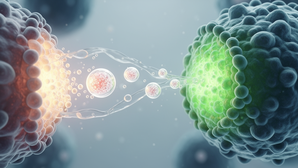
Image Credits: AI Generated
DOI: Not provided
Keywords: cellular senescence, small extracellular vesicles, paracrine senescence, evSASP, p16INK4A, IMR-90 fibroblasts, DNA damage response, gammaH2AX, Ki-67, progerin, TRF2, interleukin-6, interleukin-1beta, senescence-associated beta-galactosidase, nanoparticle tracking analysis, ultracentrifugation, cell-cycle arrest
News Source: Beatrice Stafford. (November 14, 2026). Small Extracellular Vesicles Transmit Senescence Signals in Human Cell Aging Models. Scienmag.