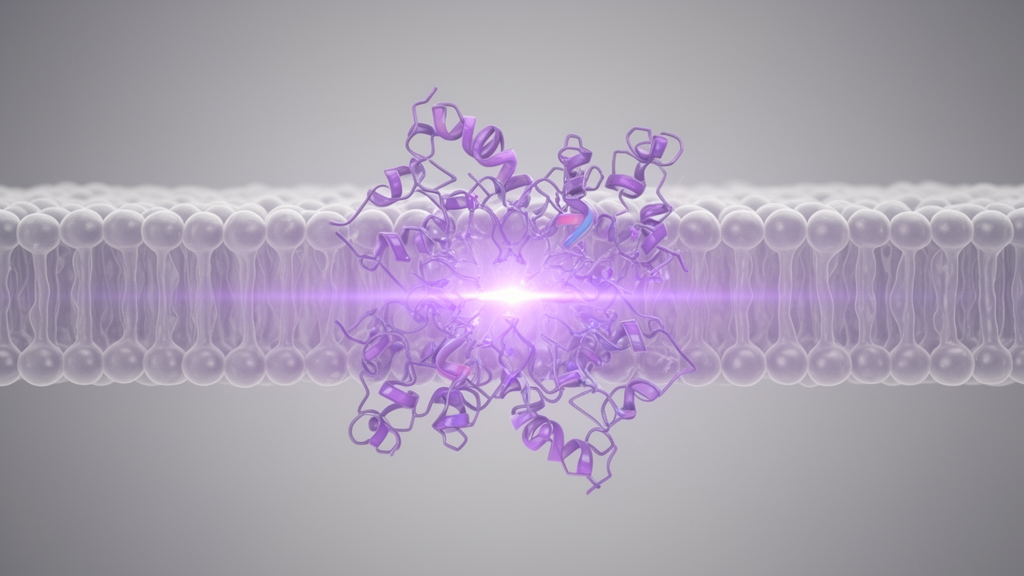
A comprehensive scientific review reveals that vertebrate non-visual opsin 5 operates across diverse organ systems through distinct, species-specific intracellular signaling cascades to regulate essential physiological processes. Documented in literature as neuropsin, OPN5 is an evolutionarily conserved G protein-coupled receptor found in mammals, birds, amphibians, and fish. While visual opsins mediate image formation, non-visual opsins govern homeostatic functions ranging from circadian photoentrainment to vascular development and neuroendocrine control. The synthesized evidence demonstrates that mammalian OPN5, designated OPN5m, functions predominantly in extra-retinal structures, including the skin, testis, and deep hypothalamic circuits, alongside selective retinal ganglion cells. Crucially, OPN5m does not share a uniform downstream pathway across vertebrates. Human receptors preferentially mobilize intracellular calcium via Gq signaling, mouse orthologs recruit Gi proteins to suppress cyclic adenosine monophosphate, avian receptors engage either G protein depending on anatomical context, and lower vertebrates primarily exploit Gq pathways to coordinate physiological responses to environmental violet light.
Structurally, OPN5 possesses the seven-transmembrane architecture of G protein-coupled receptors and binds retinal as its chromophore. OPN5m functions as a bistable opsin whose inactive 11-cis-retinal-bound and active all-trans-retinal-bound states are thermally stable and interconvertible by light, eliminating the need for enzymatic chromophore regeneration. Mammalian OPN5m exhibits peak absorbance around 380 nanometers within the violet and ultraviolet spectrum, whereas chicken OPN5m peaks near 360 nanometers. In contrast, human visual opsins absorb longer wavelengths, with cone opsins peaking between 420 and 560 nanometers, and melanopsin peaking near 480 nanometers. Violet light triggers 11-cis-retinal photoisomerization to all-trans-retinal, initiating G protein signaling, whereas subsequent orange light exposure exceeding 520 nanometers restores the inactive resting state. Notably, human and mouse OPN5m cannot bind all-trans-retinal directly due to a single amino acid substitution, requiring retinal pigment epithelium-specific 65 kDa protein expression near hypothalamic OPN5m neurons to supply external 11-cis-retinal to maintain ongoing photoreceptive function.
Intracellular signaling downstream of mammalian OPN5m diverges fundamentally between species. In cultured human cells expressing human OPN5m, ultraviolet stimulation produces transient calcium release from internal stores and activates mitogen-activated protein kinases. Mechanistic genetic knockout studies confirmed that these light-induced calcium transients depend selectively on Gq/11 proteins rather than Gi proteins, as light-evoked responses persist in Gi knockout cells but vanish in Gq/11 knockouts. Furthermore, assays monitoring G protein-coupled inwardly rectifying potassium channels revealed that ultraviolet illumination inhibits channel currents via Gq pathways, particularly through Galpha-14 coupling, demonstrating an absence of Gi involvement in cultured human cells. Conversely, mouse OPN5m selectively couples to Gi proteins in cell culture. Violet light exposure significantly reduces intracellular cyclic adenosine monophosphate by inhibiting adenylate cyclase activity. This light-dependent suppression of cyclic adenosine monophosphate confirms murine OPN5m as a functional Gi-coupled receptor, demonstrating how mammalian homologs utilize contrasting biochemical cascades to process short-wavelength light signals.
Avian OPN5m systems display remarkable functional versatility, deploying either Gi or Gq signaling cascades depending on tissue type and experimental context. In native chicken retinal ganglion cells, pineal tissue, and paraventricular hypothalamic neurons, chicken OPN5m couples primarily to Gi proteins in the presence of 11-cis-retinal, confirmed by GTP-gamma-S biochemical assays. However, when heterologously expressed in mammalian cells including HEK293T cells and mouse astrocytes, chicken OPN5m engages Gq pathways upon blue light stimulation, triggering intracellular calcium release and protein kinase C activation. This capability enabled researchers to apply avian OPN5m as an optogenetic tool to manipulate neuronal circuits and animal behavior. In quail, recordings from Xenopus oocytes and hypothalamic paraventricular neurons show that short-wavelength light stimulates calcium-activated chloride currents and rapid depolarization via Gq signaling. Conversely, canary hypothalamic OPN5m couples to Gi proteins, where RNA interference demonstrated that receptor activity suppresses thyroid-stimulating hormone beta-subunit transcription, thereby inhibiting photoperiod-induced reproductive activation.
In amphibians and ray-finned teleost fish, OPN5m homologs operate primarily through Gq-type G proteins to mediate intracellular calcium elevation. Experiments in Xenopus tropicalis, zebrafish, and medaka demonstrate light-induced calcium transients that are enhanced in the presence of Galpha-14, indicating a conserved Gq signaling mechanism across non-mammalian vertebrates. In medaka, OlOPN5m is uniquely expressed in pituitary melanotrophs among 34 examined opsins. Activation of OlOPN5m by short-wavelength ultraviolet light between 365 and 460 nanometers induces rapid calcium entry and triggers the exocytosis of melanocyte-stimulating hormone. This endocrine response promotes cutaneous melanogenesis by upregulating tyrosinase expression, establishing an autonomous non-visual endocrine pathway that protects tissues from ultraviolet damage. Furthermore, ray-finned fishes express two distinct paralogs, OPN5m and OPN5m2. Unlike OPN5m, OPN5m2 exclusively binds 11-cis-retinal and shows species-specific brain distribution: OPN5m2 is expressed in spotted gar brain but absent in zebrafish brain, while medaka lack the OPN5m2 paralog entirely from their genome.
Within the mammalian eye, OPN5m executes vital developmental and refractive regulatory functions through distinct retinal ganglion cell populations. Nguyen and colleagues discovered that light-dependent OPN5m signaling in retinal ganglion cells governs hyaloid vessel regression during postnatal ocular vascular development. Near-ultraviolet stimulation of retinal ganglion cell OPN5m lowers vitreous dopamine levels via vesicular GABA transporter and dopamine transporter phosphorylation, activating dopamine D2 receptors to suppress vascular endothelial growth factor receptor 2 activity in hyaloid endothelial cells. Furthermore, Jiang and coworkers demonstrated that OPN5 suppresses lens-induced myopia in mice by sensing violet light. Violet light exposure ameliorated lens-induced myopia in wild-type mice, but this protective effect was attenuated in conditional knockout mice lacking ocular OPN5, generated by crossing Chx10-cre with OPN5-floxed mice. This myopia suppression is accompanied by violet-light-induced upregulation of early growth response protein 1 in the retina, suggesting an essential role for violet-light-driven OPN5m activation in maintaining healthy ocular refractive development.
In mammalian skin, OPN5m is localized to hair follicle stem cells and melanocyte progenitor cells at the base of hair follicles in ear and vibrissal pad tissue. OPN5m senses ultraviolet light and stimulates melanogenesis to protect cutaneous cells from radiation damage, while ex vivo experiments show that OPN5m mediates the photoentrainment of local skin circadian clocks. At the behavioral level, OPN5-null mice exhibit impaired circadian photoentrainment and phase shifting under UVA light. However, triple-knockout mice lacking rods, cones, and melanopsin fail to entrain to UVA cycles despite expressing OPN5m, demonstrating that OPN5m expression alone is insufficient for central behavioral circadian photoentrainment and requires functional interactions with other retinal photoreceptors. Additionally, while corneal OPN5m expression facilitates wound healing, OPN5 knockout does not cause gross pigmentation defects like albinism, confirming that cutaneous OPN5m functions in dynamic homeostatic adaptation rather than establishing baseline physiological melanin production across dermal layers.
Deep within the mammalian brain, OPN5m regulates metabolism and body temperature from the hypothalamic preoptic area. Neuroanatomical studies in young and adult mice reveal that OPN5m expression is restricted to preoptic neurons, where over 95 percent are glutamatergic and positive for pituitary adenylate cyclase-activating peptide. Furthermore, over 80 percent express brain-derived neurotrophic factor mRNA, and approximately half express the heat-sensitive ion channel TrpM2, whereas prostaglandin D2 synthase is rare. Chemogenetic activation or inhibition of these OPN5m neurons significantly decreases and increases body temperature, respectively. In addition, ex vivo experiments utilizing a FRET-based cAMP sensor demonstrate that violet light illumination directly increases intracellular cyclic adenosine monophosphate in these hypothalamic cells. These results indicate that preoptic OPN5m neurons detect violet light to regulate metabolic heat generation, although whether sufficient natural ambient light reaches the preoptic area through mammalian hair, skin, and skull to drive physiological regulation remains unresolved in living animals.
In non-mammalian vertebrates, deep-brain OPN5m coordinates seasonal neuroendocrine rhythms and early larval behaviors through direct encephalic photoreception. In Japanese quail, OPN5m is expressed in neurons within the hypothalamic paraventricular organ contacting the cerebrospinal fluid. Because avian craniums allow short-wavelength light transmission, 419-nanometer light directly activates these deep neurons to stimulate thyroid-stimulating hormone release and promote testicular growth in eye-patched birds. In Xenopus laevis, XtOPN5m expression begins during embryonic stages 37 and 38, distributing across bipolar, amacrine, and ganglion cells by stage 41, where illumination induces c-Fos expression. XtOPN5m is also expressed in the larval pineal complex and caudal diencephalon, where short-wavelength light sensing induces rhythmic swimming bouts. In ray-finned fishes, retinal OPN5m is predominantly expressed in the inner nuclear layer and hypothalamic regions, while medaka OlOPN5m mediates pituitary hormone release, demonstrating that non-visual opsins across diverse vertebrate clades link deep-tissue light detection directly to essential endocrine and motor outputs.
Investigating mammalian OPN5m presents notable technical challenges that complicate precise interpretation of its in vivo functions. Retinal expression levels are exceptionally low, and researchers face a persistent scarcity of widely validated, commercially available OPN5 antibodies. Furthermore, lineage-tracing experiments using OPN5-cre crossed with Ai14 reporter mice yield tdTomato expression that persists even if OPN5 transcription occurred only transiently during embryonic development, leaving uncertainty regarding continuous expression in mature retinal ganglion and inner nuclear layer cells. Clinically, the review underscores that modern indoor environments severely limit exposure to natural violet light between 360 and 400 nanometers due to ultraviolet-filtering window glass and interior LED fixtures lacking short-wavelength spectra. This widespread deficit in violet light exposure may deprive OPN5m of necessary stimulation, correlating with the rising global incidence of myopia among children spending decreased time outdoors. Future investigations into OPN5m mechanisms may inform novel preventative therapeutics and healthier architectural lighting designs to mitigate emerging public health concerns.
Subject of Research: Physiological functions and species-specific signaling mechanisms of non-visual opsin 5
Article Title: The physiological functions of OPN5m
Article References: Ma, J., Rahaman, S. M., Ohmura, Y., & Yamanaka, A. (2025). The physiological functions of OPN5m. Eye and Vision, 12(1), Article 48. https://doi.org/10.1186/s40662-025-00467-9
Image Credits: AI Generated
DOI: 10.1186/s40662-025-00467-9
Keywords: OPN5m, neuropsin, violet light, non-visual opsins, G protein-coupled receptor, circadian photoentrainment, myopia suppression, hyaloid vessel regression, hypothalamic preoptic area, thermoregulation, melanogenesis, retinal ganglion cells, cyclic adenosine monophosphate, Gq protein signaling, Gi protein coupling, medaka pituitary, early growth response protein 1, bistable opsin
News Source: Drew Townsend. (November 14, 2026). Violet Light Receptor OPN5 Regulates Diverse Non-Visual Functions Across Vertebrates. Scienmag.