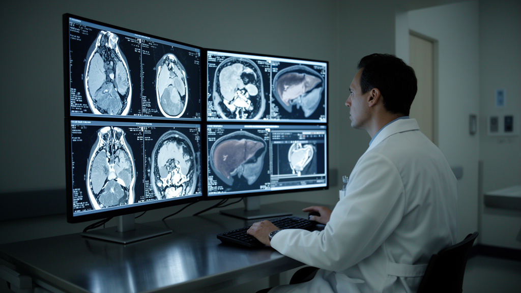
A multicenter retrospective investigation has demonstrated that integrating qualitative magnetic resonance imaging observations with deep learning and intratumoral habitat radiomics can reliably identify the aggressive macrotrabecular-massive subtype of hepatocellular carcinoma before surgical intervention while independently stratifying patient risks of postoperative disease recurrence. The research, conducted across three medical institutions and published in European Radiology Experimental, evaluated preoperative contrast-enhanced scans from 607 patients who subsequently underwent curative-intent hepatic resection. By merging computational image analyses with serological biomarkers and qualitative radiological assessments, the investigators established an interpretable fusion nomogram designed to overcome the diagnostic blind spots of conventional imaging evaluations. In validation testing across independent hospital cohorts, this multimodal scoring model achieved an area under the receiver operating characteristic curve of 0.858 for detecting the macrotrabecular-massive phenotype, significantly outperforming traditional clinical and radiological models. Furthermore, patients categorized as harboring this high-risk histological variant by the predictive nomogram exhibited markedly shortened recurrence-free survival intervals following liver resection, demonstrating that preoperative computational imaging can assist clinical teams in identifying biologically aggressive tumors prior to invasive tissue sampling.
Primary hepatocellular carcinoma represents one of the most frequently diagnosed malignant liver neoplasms worldwide and continues to exhibit high rates of recurrence following curative surgical resection. Although specific circulating proteins such as Endothelial-Specific Molecule 1 have shown promise as potential biomarkers, definitive classification has historically required invasive histopathological analysis of resected specimens, which inherently prevents personalized surgical planning or neoadjuvant considerations. Conventional radiomics approaches have attempted to address this clinical gap by extracting quantitative pixel patterns from whole tumor volumes, but standard whole-tumor analyses frequently obscure internal biological heterogeneity. In contrast, habitat radiomics isolates distinct subregions within a lesion to capture varying microenvironments related to cellular density, vascularity, and necrosis. Simultaneously, three-dimensional deep learning models capture hierarchical spatial abstractions that may escape handcrafted mathematical filters. Developing a reliable noninvasive preoperative pipeline that fuses these advanced imaging technologies with standard clinical indicators has therefore stood as a vital objective for precision hepatobiliary oncology.
The study cohort comprised 739 initial candidates diagnosed with pathologically confirmed hepatocellular carcinoma between June 2015 and January 2024 across three participating medical centers, ultimately enrolling 607 eligible patients after strict exclusion criteria. Individuals were included if they underwent gadolinium-ethoxybenzyl-diethylenetriamine pentaacetic acid enhanced magnetic resonance imaging within fourteen days prior to partial hepatectomy and possessed complete postoperative histopathological records regarding macrotrabecular-massive status. Patients were excluded if they had received preoperative local-regional therapies, exhibited distant metastatic spread or concurrent secondary malignancies, or possessed incomplete clinical data or poor-quality scans. The final cohort consisted of 474 male and 133 female participants with a mean age of 59.6 years, among whom 151 patients presented with confirmed macrotrabecular-massive hepatocellular carcinoma. A primary institution cohort of 435 patients was randomly divided in a seven-to-three ratio into a training set of 304 individuals and an internal validation set of 131 individuals. The remaining 172 patients from the second and third medical institutions formed an entirely independent external validation cohort. When multiple hepatic lesions were detected, analysis was standardized by selecting the largest lesion for segmentation.
Histological examination served as the reference standard, conducted independently by two senior pathologists with eight years of specialized experience who adhered strictly to the 2019 World Health Organization classification criteria. Under these pathological definitions, a macrotrabecular architecture was identified whenever tumor cell cords exceeded six cells in thickness, and lesions were classified as macrotrabecular-massive positive if this specific structural architecture occupied more than fifty percent of the total examined tumor volume. The interobserver agreement between the two evaluating pathologists across all 607 patients reached an excellent level, reflected by a Cohen kappa coefficient of 0.92. Magnetic resonance imaging acquisitions were performed across three different commercial 3-Tesla scanners equipped with sixteen-channel phased-array torso coils, specifically utilizing Siemens MAGNETOM Verio, Philips Achieva, and GE Discovery MR750 systems. The liver-specific hepatobiliary contrast agent Gd-EOB-DTPA was administered through high-pressure intravenous injection at a flow rate of 1 to 2 milliliters per second at a standard dose of 0.025 millimoles per kilogram. Dynamic acquisitions utilized voxel-interpolated breath-hold fat-suppressed T1-weighted sequences capturing arterial, portal venous, and hepatobiliary phases at 30 seconds, 60 seconds, and 20 minutes post-injection.
Two independent radiologists with three and five years of clinical experience retrospectively evaluated qualitative features based on the Liver Imaging Reporting and Data System, reaching high inter-reader reproducibility with kappa values ranging from 0.86 to 0.91. Evaluated characteristics included infiltrative appearance, intratumoral blood products, fat within the mass, nonrim and rim arterial phase hyperenhancement, peripheral and nonperipheral washout patterns, capsule enhancement types, arterial peritumoral enhancement, intratumoral arteries, substantial necrosis exceeding twenty or fifty percent, satellite nodules, central scars, and peritumoral hypointensity during the hepatobiliary phase. To ensure volumetric precision across multi-phase scans, three-dimensional volumes of interest for 353 lesions were manually contoured across arterial, portal venous, and hepatobiliary images using 3D Slicer software to train an automated nnU-Net deep learning framework. The nnU-Net architecture was trained on 282 lesions and validated on 71 lesions using a full-resolution configuration. When deployed on the remaining 254 lesions, nnU-Net achieved Dice similarity coefficients of 0.891 for arterial phase, 0.883 for portal venous phase, and 0.932 for hepatobiliary phase segmentations. All automated masks were subsequently reviewed and adjusted by a senior radiologist with twelve years of experience.
Intratumoral heterogeneity was quantitatively mapped by extracting habitat radiomics features from the segmented volumes across all three contrast phases following spatial normalization to isotropic voxels measuring one cubic millimeter. Image voxel intensities were discretized using a fixed bin width of five to compute stable texture matrices. To define biological subregions without user bias, unsupervised K-means clustering was executed across cluster numbers ranging from two to eight, with the Calinski-Harabasz index identifying three distinct clusters as the optimal configuration. Using PyRadiomics software, 1,132 radiomics features were extracted from each individual habitat subregion across original scans, Laplacian-of-Gaussian spatial filterings with sigma values from 2.0 to 5.0 millimeters, and eight-directional wavelet decompositions. In total, 3,396 quantitative habitat features were obtained per single phase for each patient. Combined-phase feature pools integrating arterial, portal venous, and hepatobiliary acquisitions were normalized and selected using the FeAture Explorer platform to train support vector machine classifiers that generated a composite habitat radiomics score.
Deep learning representations were simultaneously extracted using a three-dimensional Swin Transformer neural network architecture pretrained on the Kinetics video dataset and implemented within PyTorch. To minimize scanner-induced signal variations, volumetric image inputs were scaled into uniform pixel intensity ranges between zero and one, complemented by built-in Layer Normalization layers that provided feature-wise normalization during model optimization. Data augmentation pipelines incorporated random spatial resizing, intensity scaling, three-dimensional rotations, flips, and affine transformations to prevent network overfitting during training sessions spanning 200 to 500 epochs. Optimization was conducted using the Adam algorithm with an initial learning rate of 0.0001, scheduled learning rate decays of tenfold every seven epochs, a mini-batch size of 16, and focal loss functions tailored to mitigate class imbalances. Extracted deep representations across the three dynamic phases were integrated into combined-phase feature sets and modeled via an autoencoder classifier to compute a continuous deep learning score. To provide clinical interpretability, the investigators utilized weighted class activation mapping, producing spatial heatmaps that confirmed the deep neural network prioritized active, hypervascular peripheral tumor areas during inference.
Model construction proceeded by first establishing a baseline clinical-radiological model through univariable and multivariable stepwise logistic regression guided by the Akaike information criterion. In this baseline formulation, serum alanine aminotransferase, alpha-fetoprotein, maximum tumor diameter, fat within the mass, capsule appearance, and peritumoral hypointensity on hepatobiliary phase imaging were identified as significant parameters. The baseline clinical-radiological model achieved areas under the curve of 0.801 in the training cohort, 0.651 in the internal validation cohort, and 0.739 in the external testing cohort. Subsequently, investigators developed the comprehensive fusion nomogram by entering all candidate variables into a multivariable backward logistic regression. Five variables emerged as statistically significant independent predictors of macrotrabecular-massive hepatocellular carcinoma: habitat radiomics score with an odds ratio of 3.561, deep learning score with an odds ratio of 3.060, serum alpha-fetoprotein with an odds ratio of 3.569, alanine aminotransferase with an odds ratio of 2.419, and the presence of fat within the mass, which showed an inverse protective association reflected by an odds ratio of 0.149.
The resulting multimodal fusion nomogram demonstrated superior diagnostic accuracy compared to the standard clinical-radiological approach across every evaluation cohort. The nomogram yielded an area under the receiver operating characteristic curve of 0.897 with a 95% confidence interval of 0.854 to 0.940 in the training set, 0.757 with a 95% confidence interval of 0.674 to 0.827 in the internal validation set, and 0.858 with a 95% confidence interval of 0.797 to 0.906 in the external testing cohort. DeLong statistical testing confirmed that these diagnostic gains were statistically significant over the clinical-radiological model across training, internal validation, and external testing groups, yielding p-values of less than 0.001, 0.017, and 0.015, respectively. Calibration curves verified close agreement between nomogram predictions and true histological outcomes across all centers, while decision curve analysis indicated positive net clinical benefit across relevant intervention thresholds. Shapley additive explanations further clarified the internal logic of the model, demonstrating that elevated radiomics scores, elevated deep learning scores, high alpha-fetoprotein levels, and high alanine aminotransferase concentrations contributed positively toward a predicted macrotrabecular-massive diagnosis, whereas the presence of fat within the tumor consistently exerted a negative predictive weight.
Prognostic evaluation extended beyond diagnostic classification to analyze recurrence-free survival among 536 enrolled patients who maintained complete follow-up through December 2024, during which 231 disease recurrence events were documented. Across all datasets, patients classified as harboring macrotrabecular-massive tumors by the nomogram experienced significantly shorter median recurrence-free survival compared to those predicted as negative: 18.9 versus 39.3 months in the training set, 26.3 versus 40.5 months in internal validation, and 30.0 versus 46.4 months in external testing cohorts. Multivariable Cox proportional hazards regression demonstrated that elevated alpha-fetoprotein with a hazard ratio of 2.76 and nomogram-predicted macrotrabecular status with a hazard ratio of 1.72 operated as independent prognostic risk factors for tumor recurrence. Despite these robust findings, the authors highlighted important limitations, including the retrospective nature of the study, the lack of dedicated scanner harmonization across heterogeneous imaging hardware, computational demands that currently prevent real-time intraoperative deployment, and the absence of long-term overall survival metrics. Nevertheless, the study demonstrates that noninvasive fusion nomograms combining spatial habitat radiomics, deep learning representations, and standardized radiological observations provide clinical value for preoperative staging and post-resection patient monitoring.
Subject of Research: Preoperative prediction of macrotrabecular-massive hepatocellular carcinoma and recurrence-free survival using multimodal MRI analysis
Article Title: Interpretable prediction of macrotrabecular-massive HCC and recurrence-free survival by integrating MRI LI-RADS features with deep learning habitat radiomics
Article References: Zhang, M., Wu, Q., Zhu, M., Zhang, T., Gu, C., Gu, W., Du, M., Shen, H., Hu, C., Yang, Y., Wang, X., & Yu, Y. (2026). Interpretable prediction of macrotrabecular-massive HCC and recurrence-free survival by integrating MRI LI-RADS features with deep learning habitat radiomics. European Radiology Experimental, 10(1), Article 121. https://doi.org/10.1186/s41747-026-00795-y
Image Credits: AI Generated
DOI: 10.1186/s41747-026-00795-y
Keywords: Hepatocellular Carcinoma, Macrotrabecular-Massive HCC, Magnetic Resonance Imaging, Habitat Radiomics, Deep Learning, Swin Transformer, LI-RADS, Recurrence-Free Survival, Gd-EOB-DTPA, Tumor Heterogeneity, Multivariate Logistic Regression, Cox Proportional Hazards, Alpha-Fetoprotein, Alanine Aminotransferase, nnU-Net Segmentation, Nomogram, Decision Curve Analysis
News Source: Nathaniel Bowman. (November 14, 2026). MRI Nomogram Predicts Aggressive Liver Cancer Subtype and Postoperative Recurrence. Scienmag.