Researchers have engineered an optogenetic tool termed ASAP that acutely inhibits cellular autophagy within five minutes of blue-light exposure, enabling the precise dissection of rapid degradation kinetics and identifying previously unrecognized substrates under basal conditions. Developed by an investigative team led by Christina G. Towers and Payel Mondal, the system overcomes long-standing barriers that historically obscured the immediate consequences of halting autophagic turnover. By providing rapid, reversible control over autophagosome-lysosome fusion using light, ASAP allows researchers to track constitutive degradation processes with unprecedented temporal resolution.
Prior to ASAP, investigators studying autophagy perturbation relied predominantly on chemical inhibitors like bafilomycin-A1 or genetic techniques utilizing CRISPR-Cas9 and doxycycline-inducible cassettes. While bafilomycin-A1 acts as a lysosome inhibitor, experimental comparisons showed it requires one to two hours to initiate detectable accumulation of LC3-II and up to four hours to inhibit autophagy. Furthermore, chemical washout fails to reverse autophagy blockade even after twenty-four hours in drug-free medium.
To overcome these limitations, the researchers engineered ASAP by repurposing a dominant-negative variant of syntaxin-17, known as STX17ΔNTD, which lacks its amino-terminal domain. To place this dominant-negative effector under photochemical control, the team fused STX17ΔNTD to a light-oxygen-voltage hybrid domain, hLOV1, engineered to cage a cleavage recognition sequence for tobacco etch virus protease, termed tevS. The construct incorporates an amino-terminal immunoglobulin kappa leader sequence and a platelet-derived growth factor receptor transmembrane domain, anchoring the chimeric polyprotein to the endoplasmic reticulum. A ribosomal skipping p2A sequence was inserted between the caged fusion protein and the protease, ensuring stoichiometric expression of both ASAP and the protease from a single promoter.
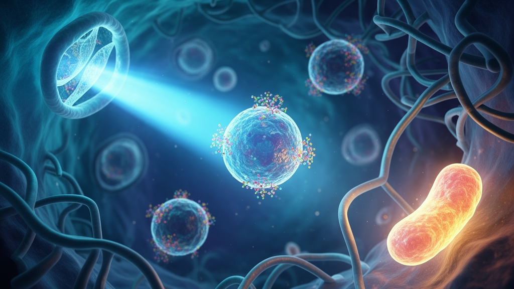
The molecular architecture of ASAP guarantees spatial and functional sequestration under dark conditions, leaving basal autophagy uninhibited until an optical trigger is applied. In the dark, unactivated hLOV1 shields the internal tevS recognition sequence from proteolytic access while the transmembrane segment tethers STX17ΔNTD to the endoplasmic reticulum, away from autophagosomes. When illuminated with pulsed 450-nanometer blue light delivered at intervals of ten seconds on and five seconds off, hLOV1 undergoes an instantaneous conformational change that uncages the tevS site. The co-expressed tobacco etch virus protease rapidly cleaves the accessible site, releasing the myc-tagged STX17ΔNTD fragment into the cytoplasm. Once liberated, the dominant-negative peptide diffuses through the cytosol and binds to autophagosomes, preventing their fusion with lysosomes and arresting autophagic flux within five minutes of light exposure.
The researchers validated the operational kinetics of ASAP across diverse cultured human cell models, including HEK293T cells, HCT116 colorectal carcinoma cells, and NCI-H292 lung carcinoma cells. Immunofluorescence microscopy and subcellular fractionation confirmed that intact ASAP remained localized to the endoplasmic reticulum in the dark, overlapping with SERCA2 without appreciable trafficking to the plasma membrane. Upon blue-light illumination, immunoblotting detected cleaved STX17ΔNTD within five minutes, accompanied by progressive accumulation of lipidated LC3-II and a transient rise in the autophagy receptor p62, also known as SQSTM1. Co-treatment with light and bafilomycin-A1 produced no additive increase in LC3-II accumulation, confirming that ASAP specifically blocks autophagic degradation.
A distinctive operational advantage of ASAP is its rapid reversibility upon cessation of light stimulation, driven by endogenous cellular clearance mechanisms. When illuminated cells were returned to complete darkness, immunoblotting revealed that the liberated STX17ΔNTD fragment exhibited a cellular half-life of approximately thirty minutes, restoring baseline LC3-II levels within one hour. Experimental addition of the proteasome inhibitor MG132 prevented this recovery, demonstrating that ASAP reversibility depends directly on proteasomal degradation of the unstable cleaved effector fragment. Control experiments confirmed that the optical dosage of 450-nanometer light, delivered at an irradiance of 0.6 to 0.7 milliwatts per square centimeter, caused neither reactive oxygen species accumulation nor caspase-3/7 apoptotic activation in untransfected cells. Prolonged intermittent illumination revealed that ASAP-expressing cells manifested noticeable growth defects starting at twenty-four hours, whereas cells subjected to continuous bafilomycin-A1 required three days to show comparable growth inhibition.
Taking advantage of the rapid temporal control afforded by ASAP, the authors coupled acute light inhibition to multiplexed tandem mass tag mass spectrometry to profile the human cellular proteome. By comparing HEK293T cells expressing ASAP against control constructs after five or thirty minutes of blue-light exposure, the team quantified 5,219 high-confidence proteins across all subcellular compartments under undisturbed basal conditions. Unlike previous proteomic investigations that enforced prolonged genetic ablation or relied on harsh stressors, this approach identified substrates that accumulate immediately upon pathway arrest, distinguishing constitutive turnover targets from indirect downstream adaptations. Statistical analysis using Welch-corrected t-tests and Benjamini-Hochberg false-discovery rate adjustments revealed significant accumulation of known autophagy adaptors, including GABARAP and STX17, within thirty minutes.
Proteomic profiling and follow-up immunoblotting demonstrated that mitochondrial quality control constitutes one of the most prominent constitutive degradation pathways operating under unperturbed growth conditions. Within five minutes to three hours of ASAP activation, proteins residing on the outer mitochondrial membrane accumulated rapidly and sustainably, while mitochondrial matrix proteins remained stable over the same timeframe. Notably, ubiquitin-independent mitophagy receptors embedded in the outer membrane, including BNIP3, BNIP3L (also designated NIX), and FUNDC1, exhibited pronounced protein accumulation without changes in their corresponding messenger RNA levels. Conversely, canonical ubiquitin-dependent mitophagy receptors such as OPTN, TAX1BP1, and NDP52 showed minimal accumulation during acute inhibition timepoints.
The most unexpected discovery emerging from the proteomic screen was that nucleoporins, the constituent structural proteins of the nuclear pore complex, undergo constitutive autophagic degradation in mammalian cells. Quantitative immunoblotting confirmed that nucleoporins NUP54, NUP88, and NUP214 accumulated significantly in whole-cell lysates within five minutes of light exposure without increases in gene transcription. Pre-treatment with the translation inhibitor cycloheximide abolished this cytoplasmic accumulation during illumination, proving that accumulating nucleoporins represent newly synthesized polypeptides rather than disassembly products of preexisting nuclear envelope complexes.
Investigating the biochemical mechanism governing mammalian nucleophagy revealed that specific nucleoporins possess evolutionarily conserved LC3-interacting regions that directly mediate recognition by the core autophagy machinery. Sequence analysis identified two predicted canonical LC3-interacting motifs within human NUP214 and one conserved motif within human NUP88, matching the aromatic-hydrophobic consensus sequence necessary for binding ATG8-family proteins. Co-immunoprecipitation assays demonstrated that wild-type NUP214 and NUP88 physically interact with Flag-tagged and endogenous LC3 along with associated nucleoporins NUP358 and NUP62. Site-directed mutagenesis mutating critical aromatic and aliphatic residues to alanine revealed that NUP214 functions as the primary autophagy receptor; disrupting both LC3-interacting regions in NUP214 abolished its binding to LC3 and simultaneously prevented co-precipitation of non-receptor nucleoporins NUP358 and NUP62. While NUP88 point mutations diminished its direct interaction with LC3, residual complex binding persisted, demonstrating that NUP214 acts as the principal adaptor directing pre-assembled cytoplasmic nucleoporin subcomplexes into autophagic degradation pathways.
Immunofluorescence imaging revealed that the number of DCP1A-positive processing bodies increased within ten minutes of ASAP activation, displaying marked spatial colocalization with cytoplasmic nucleoporins including NUP358, whereas G3BP-positive stress granules were not induced. Despite its experimental power, the authors note several limitations of ASAP, including reliance on transient plasmid transfection, subtle dark-state basal leakage at elevated expression levels, potential off-target effects related to mitochondrial STX17 localization, and the inability of mass spectrometry to detect small lipidated proteins. Nevertheless, ASAP provides an agile optogenetic framework to manipulate autophagy acutely across diverse biological models, illuminating real-time proteostatic surveillance mechanisms.
Subject of Research: Rapid optogenetic inhibition of autophagy and identification of constitutive nucleoporin degradation
Article Title: The optogenetic tool ASAP acutely inhibits autophagy
Article References: Mondal, P., Cyril, A., Mamriev, D., Parham, L., Wierzbicki, I., Shen, C., Doglioni, G., Dufrense, S., Komarla, A., Gonzalez, D. J., D’Angelo, M. A., & Towers, C. G. (2026). The optogenetic tool ASAP acutely inhibits autophagy. Nature Cell Biology. https://doi.org/10.1038/s41556-026-02124-2
Image Credits: AI Generated
DOI: 10.1038/s41556-026-02124-2
Keywords: autophagy inhibition, optogenetics, ASAP tool, STX17, nucleoporins, nuclear pore complex, NUP214, NUP88, NUP358, LC3-interacting regions, mitophagy, processing bodies, bafilomycin-A1, tandem mass tag proteomics, HEK293T cells, annulate lamellae, hLOV1 domain
News Source: Drew Townsend. (November 12, 2026). Optogenetic Tool ASAP Shuts Down Cellular Autophagy Within Five Minutes. Scienmag.