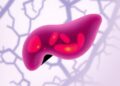
Credit: Prof. Ichiro Masai
There are many different structures in our eyes that work in conjunction to allow us to see. These structures are strikingly similar between different species, from zebrafish to humans. The growth of ocular tissues must be tightly controlled in order to maintain the correct eye size and shape that allow us to see. This tight regulation has intrigued developmental biologists for decades.
The lens of the eye focuses incoming light on the retina, which then converts the light into electrical signals allowing us to see. Two distinct cell types comprise the lens: epithelial cells, which cover the front, or anterior, portion of the lens, and fiber cells, which populate the back, or posterior, portion. It has been shown that epithelial cells proliferate in the anterior half of the lens and move towards the posterior half, differentiating, or transforming, into fiber cells when they reach the equator between the two halves. In order to elucidate the underlying mechanisms that drive this movement, the Developmental Neurobiology Unit at the Okinawa Institute of Science and Technology Graduate University (OIST), led by Prof. Ichiro Masai, employed time-lapse imaging techniques to observe real time lens development in zebrafish. Their results were recently published in Development.
Dr. Toshiaki Mochizuki, along with OIST students Yi-Jyun Luo and Hsieh-Fu Tsai, developed a technique that allowed them to track the movement and development of lens epithelial cells in a live zebrafish embryo in real-time. The researchers used genetically engineered zebrafish that contain two different fluorescent proteins, mCherry-zGem and GFP-histones, that would fluoresce, or emit light to produce a color, at different points in the cell cycle. During the initial G1 phase of the cell cycle, only GFP is expressed, giving the cells a green appearance under a confocal microscope. As the cell then enters the S phase, during which DNA replication occurs, expression of the mCherry-zGem protein begins, changing the color from green to yellow. As the expression of the mCherry-zGem proteins increases in the subsequent G2 and M phases, the color shifts to red and deep red, respectively. Cells that do not enter the cell cycle, and stay in the quiescent G0 phase, never express the mCherry-zGem protein and thus remain green throughout the experiment. The color changes, or lack thereof, effectively allow the scientists to monitor the phases of the cell cycle of each epithelial cell.
The OIST researchers then analyzed the time lapse images from the zebrafish lens to reveal that the epithelial cells segregate into dividing cells and non-dividing cells. Dividing cells enter the cell cycle and thus display a change in color, while non-dividing cells remain green throughout the duration of the experiment. The scientists saw that groups of non-dividing cells would move as a cluster in a spiral-like pattern following division of neighboring cells. This division would prompt the non-dividing cells to move towards the equator of the lens, towards differentiation into fiber cells.
Additionally, the Developmental Neurobiology Unit discovered that the movement of cells in the lens also appeared to be regulated by two related proteins: E-cadherin, expressed in lens epithelial cells, and N-cadherin, expressed in lens fiber cells. These proteins exert opposite forces on neighboring cells, with E-cadherin exerting a trapping force and N-cadherin exerting a pulling force. Together, E-cadherin and N-cadherin also help regulate cell movement through the modulation of the lens epithelial cells' adhesion and tension.
"I'm very proud that our group was able to develop a technique that allowed us to observe these cells in a living zebrafish over a long period of time", explained Masai, "This is the first time that the growth of individual lens epithelial cells has been tracked over such a long period of time. This research has allowed us to determine the factors responsible for the regulation of eye development. Without these factors, correct eye development would not be possible!"
###
Media Contact
Kaoru Natori
[email protected]
@oistedu
http://www.oist.jp/