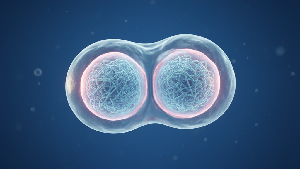
A research team led by scientists at Kyushu University and the National Institute of Genetics has discovered that the cell nucleus undergoes a pronounced, transient softening during the two-cell stage of mouse embryonic development. This structural transformation was found to be an integral mechanical driver of major zygotic gene activation, an essential developmental milestone in which approximately two thousand genes activate simultaneously to kickstart embryonic progression. Reporting their experimental findings in the journal Nature, the investigators established that mechanical deformation of the nucleus is indispensable for subsequent development, resolving a longstanding question regarding the biological role of nuclear physical properties at the very origin of life.
In eukaryotic biology, the nucleus houses the cellular genome and is physically bounded and stabilized by the nuclear envelope. The structural integrity and rigidity of this envelope are modulated across a cell’s lifecycle depending on its specialized role and physiological state, and failures in maintaining envelope architecture often precipitate cellular dysfunction. Abnormal nuclear shape is used for histological diagnosis and characterization of cancer. Some cancer cells have soft nuclei, which in theory enables them to leave their environment and travel to other parts of the body in a phenomenon called metastasis.
Despite widespread recognition of nuclear architecture in differentiated cells and disease states, the biophysical dynamics of the nucleus during the initial hours following fertilization have historically remained uncharacterized. Co-lead investigator Professor Kei Miyamoto of Kyushu University’s Faculty of Agriculture noted that while nuclear structure is known to govern essential cellular behaviors, little empirical data previously existed regarding mechanical state changes during the earliest cleavage cycles. Seeking to understand how nuclear stiffness modulates initial embryogenesis, the collaborative group investigated the physical transitions occurring within the first forty-eight hours of mammalian life, spanning the progression from single-cell zygote to the four-cell embryonic stage.
To overcome these experimental constraints, the laboratory of co-lead author Associate Professor Yuta Shimamoto at the National Institute of Genetics engineered a micro-invasive, nanocapillary-based measurement platform capable of quantifying nuclear rigidity without destroying living cells. The apparatus introduces a delicate glass micropipette, engineered with a diameter substantially smaller than the nucleus itself, directly into the blastomere until it contacts the nuclear surface. By applying calibrated micro-suction through the capillary, the system induces localized surface stretching and deformation, translating mechanical displacement into direct measurements of nuclear stiffness.
Applying this micro-mechanical platform, the researchers tracked nuclear rigidity following fertilization, during which the individual sperm and egg pronuclei fuse into a single diploid nucleus before dividing into two distinct cells. Measurements revealed that the newly formed nucleus exhibits high structural stiffness throughout the initial one-cell zygotic stage. However, following the very first cleavage division into the two-cell stage, the nucleus undergoes a striking transition, becoming considerably softer, plastic, and visibly deformed. This pliancy is strictly transient: as cleavage continues and the embryo divides into the four-cell stage, the nuclei regain their structural rigidity, returning to baseline stiffness levels comparable to the original one-cell state.
Molecular and biochemical dissections demonstrated that this temporary mechanical softening is driven by targeted structural remodeling of the nuclear lamina. Specifically, the researchers observed that Lamin B1, a foundational architectural protein that reinforces the structural scaffold of the nuclear envelope, is selectively cleared from the envelope during the two-cell stage. The temporary degradation and clearance of Lamin B1 directly induce the reduction in mechanical resistance, allowing the nuclear surface to become pliable. Cells whose nuclei did not soften at the two-cell stage were considerably less likely to develop further.
The developmental necessity of this mechanical transition is linked directly to major zygotic genome activation, commonly abbreviated as ZGA. During ZGA at the two-cell stage, over two thousand silent genes are expressed almost concurrently. The investigators demonstrated that the physical loosening and deformation of the nucleus are functionally coupled to this transcriptional burst, facilitating chromatin spatial rearrangement and promoting the coordinated expression of essential embryonic programs.
High-resolution imaging and locus-tracking experiments revealed that nuclear softening permits extensive three-dimensional spatial reorganization of gene loci within the nucleoplasm. In stiffened control nuclei, chromatin loci experienced constrained mobility, restricting critical developmental genes from shifting into transcriptionally favorable positions. Conversely, the pliable envelope of the softened nucleus permitted active transcription sites to mobilize adjacent to the softened nuclear envelope. In embryos where nuclear softening was mechanically or biochemically suppressed, active DNA transcription of ZGA genes near the nuclear periphery was markedly inhibited, demonstrating that mechanical compliance directly dictates transcriptional competence.
These experimental results demonstrate that physical mechanics act as a functional regulator of genomic transcription and embryogenesis rather than serving as passive structural consequences. Because the study focused on mouse embryonic models, the authors noted that future investigations must examine whether equivalent transient nuclear softening governs early embryonic development across other mammalian species, including humans.
Beyond foundational developmental biology, the authors highlighted tangible avenues for applied translational research. Professor Miyamoto expressed hope that in the future, this will lead to a better way of evaluating viable embryos in procedures such as IVF. Additionally, the team noted that the nanocapillary mechanics platform and the insights into transient envelope regulation could provide novel analytical tools for investigating nuclear malleability, lamina regulation, and invasive migration in metastatic cancer cells.
Structural visualization through advanced microscopy and three-dimensional reconstruction provided detailed mechanistic insight into how this mechanical softening facilitates gene expression during the two-cell stage. In two-cell mouse embryos where the twin nuclei were imaged in fluorescent green, three-dimensional renderings of individual nuclei highlighted a noticeably deformed nuclear surface rendered in blue. This distinct topological distortion serves a direct functional purpose by altering internal spatial architecture. Specifically, the inward and outward folding of the pliable nuclear boundary allows developmental gene loci displaying active transcription to physically approach RNA polymerase complexes. By drawing transcriptionally primed chromatin into close proximity with transcriptional machinery concentrated near the nuclear periphery, the mechanical deformation ensures that critical developmental sequences are efficiently transcribed into messenger RNA, visualized in red, which is subsequently exported and translated into essential embryonic proteins.
Beyond its observational utility, the experimental nanocapillary system represented a significant methodological advance because it enabled the investigators not only to quantify but also to directly manipulate the physical compliance of embryonic nuclei in real time.
Kyushu University has submitted patent applications under PCT/JP2026/20038 covering nuclear shape-based embryo assessment methods, designating Kei Miyamoto, Masahito Tanaka, and Yuta Shimamoto as inventors.
Subject of Research: Transient nuclear softening and zygotic gene activation during early mouse embryonic development
Article Title: The softening of an embryo’s nucleus after the very first division is critical for further development
Article References: The softening of an embryo’s nucleus after the very first division is critical for further development. Original publication (Bibliographic details unavailable.)
Image Credits: AI Generated
DOI: Not provided
Keywords: mouse embryonic development, two-cell stage, zygotic gene activation, nuclear softening, nanocapillary measurement system, Lamin B1, nuclear envelope, Kei Miyamoto, Yuta Shimamoto, Kyushu University, National Institute of Genetics, in vitro fertilization, DNA transcription, nuclear stiffness, embryo viability, cancer metastasis, histological diagnosis
News Source: Juliet Wilcox. (November 11, 2026). Mouse Embryo Nuclei Soften After First Division to Enable Gene Activation. Scienmag.