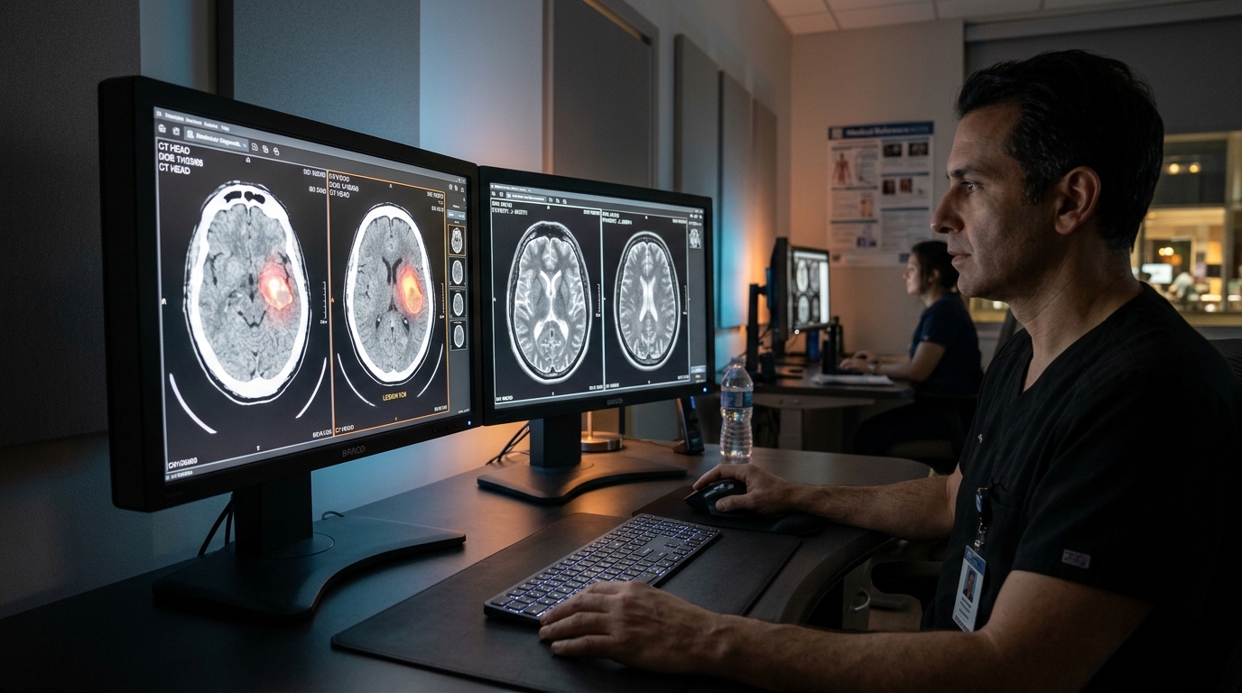
A comprehensive systematic review of 180 peer-reviewed studies published between 2015 and 2025 has established that artificial intelligence systems, particularly those utilizing deep learning, demonstrate consistently high performance in cancer detection and prognostic modeling across various imaging modalities and tumor types. While early research focused heavily on convolutional neural networks for basic pattern recognition, the field has evolved toward sophisticated architectures that integrate imaging data with clinical and genomic information, enhancing the precision of oncological assessment and individualized treatment planning.
The study selection process followed rigorous PRISMA 2020 guidelines to identify original research articles applying artificial intelligence to major oncological imaging tasks. The researchers synthesized data from studies encompassing diverse modalities, including computed tomography (CT), magnetic resonance imaging (MRI), positron emission tomography (PET), mammography, histopathology, dermoscopy, and endoscopy. Each investigation was evaluated for methodological quality using the QUADAS-AI framework, which assesses patient selection representativeness, index test development, reference standard validity, and overall flow and timing of the study. By analyzing this breadth of evidence, the reviewers aimed to characterize the current state of AI adoption in oncology, evaluate reported performance metrics, and identify significant methodological limitations that must be addressed to facilitate broader clinical translation.
Diagnostic accuracy reported across these investigations was notably robust, with several high-quality cohorts achieving area under the receiver operating characteristic curve (AUC) values exceeding 0.90 for cancer detection. In the specific context of whole-slide histopathology, certain curated datasets enabled models to reach an AUC of 0.99, demonstrating the capacity for high-precision identification of metastases. Similarly, CT and MRI-based systems showed strong screening and diagnostic performance in large-scale studies, particularly when subjected to multicenter validation. These results underscore the potential for AI to augment existing diagnostic pipelines, potentially reducing the workload pressure on radiology and pathology services while maintaining high standards for patient care and early disease intervention.
The review also detailed the distinct performance characteristics across specific clinical domains. In breast cancer screening, mammography-based deep learning models reported AUC values between 0.89 and 0.96, while lung cancer screening studies utilizing 3D convolutional neural networks trained on over 42,000 CT scans achieved an AUC of 0.94. For prostate cancer, multiparametric MRI applications provided automated lesion localization and Gleason grade prediction, which are critical for comprehensive clinical assessment. Meanwhile, in the neuro-oncology field, benchmark datasets such as BRATS facilitated the advancement of U-Net and V-Net architectures, with more recent transformer-based models like UNETR further improving the accuracy of 3D tumor segmentation and characterization.
Beyond detection, the integration of radiomics has provided quantitative imaging biomarkers that link imaging phenotypes with molecular biology and prognosis. Handcrafted feature extraction allowed researchers to predict tumor phenotypes, survival outcomes, and potential responses to immunotherapy in patients with lung cancer and glioblastoma. The review identified these radiomic signatures as essential tools in modern precision oncology, as they transform medical images into data-rich sources for molecular inference. When combined with deep learning, these hybrid modeling approaches have demonstrated improved predictive performance, suggesting that integrating disparate biological data can yield more reliable insights into tumor behavior and treatment response than single-modality models alone.
Technological trends identified in the review point toward a transition from simple classification tasks to complex multimodal and foundation model architectures. By integrating imaging, laboratory data, and genomic profiles, researchers are developing more holistic representations of oncological status. Vision transformers and self-supervised pretraining methods have emerged as particularly influential in enhancing representation learning, especially for volumetric and cross-domain data. These methodological advancements reduce dependence on large, manually labeled datasets, which have traditionally been a bottleneck in the training and generalizability of medical AI systems across diverse institutions and scanners.
Methodological quality assessment using the QUADAS-AI framework revealed that the majority of studies were of low risk of bias, characterized by clearly documented development pipelines and the use of gold-standard reference methods such as histopathology or expert consensus. However, the reviewers identified persistent limitations in reporting standards, particularly concerning patient selection, data flow, and timing. For instance, approximately 20 percent of studies lacked clear documentation of recruitment methods or exclusion criteria, and a significant portion failed to adequately describe the temporal alignment between image acquisition and the reference standard. These reporting gaps underscore a need for greater transparency and consistency in the publication of AI-driven research in medical journals.
Validation strategies remain a critical variable in the clinical applicability of these systems. The analysis revealed that only 21 percent of the included studies performed external validation using independent datasets, and only 14 percent incorporated multicenter datasets. While retrospective validation is efficient for model development, the review cautions that such studies may be susceptible to selection bias and limited demographic diversity. The authors emphasize that prospective, multicenter trials are the necessary next step to verify the robustness of AI systems in routine clinical workflows and to ensure they perform reliably across different populations and technical environments.
Explainability and clinical transparency continue to be central themes in the ongoing evolution of AI in oncology imaging. Saliency mapping and feature attribution techniques have been utilized to improve clinician trust by visualizing the diagnostic basis of algorithmic predictions. Regulatory science frameworks are also increasingly emphasizing structured reporting standards such as CLAIM to ensure that clinical deployments are safe, effective, and consistent. Federated learning approaches were noted for their ability to mitigate privacy and data-sharing constraints, allowing for collaborative model training across institutions without the need for centralized patient data, which is an essential requirement for building large-scale, reliable models.
Implementation challenges represent the final frontier for AI-assisted oncology imaging. Although the technology has shown promise in controlled settings, integrating these tools into picture archiving and communication systems (PACS) and existing electronic health record workflows requires overcoming significant medico-legal, technical, and process-oriented hurdles. The review notes that deployment must occur without increasing the reporting burden on clinicians or introducing workflow complexities. Ongoing monitoring of system performance is also essential, as models may require periodic updates to remain effective against the backdrop of evolving scanner technologies and changing clinical protocols over time.
Looking forward, the research landscape is shifting toward the implementation of large-scale foundation models and more tightly coupled molecular-oncology frameworks. The ability to accurately predict responses to therapies, such as neoadjuvant chemoradiotherapy in rectal cancer, without excessive manual intervention highlights the practical utility of these advanced architectures. Future research priorities identified by the review include the establishment of robust, outcome-based clinical endpoints, the adoption of federated multicenter validation strategies, and the continued development of explainability frameworks to ensure transparency. As these elements mature, they are expected to have a sustained impact on precision oncology and the overall quality of routine cancer care provided globally.
In conclusion, artificial intelligence has established a strong technical foundation for cancer detection and prognostic modeling, with consistent performance verified across diverse imaging modalities. The highest clinical evidence currently supports its utility as an adjunctive tool in breast and lung cancer screening, metastatic detection in digital pathology, and the classification of dermatologic and endoscopic lesions. While the field has demonstrated significant potential, sustained clinical adoption depends on the rigorous application of prospective, multicenter validation, standardized reporting, and the successful navigation of complex regulatory and workflow integration requirements. Continuous efforts in these areas will be vital for realizing the long-term benefits of these innovative imaging technologies in clinical oncology.
Subject of Research: Systematic review of artificial intelligence applications in oncology diagnostic imaging.
Article Title: Clinical validation of artificial intelligence for cancer detection prognosis and analysis across imaging modalities and tumor types
Article References: Mohamed, N. A. R. (2026). Clinical validation of artificial intelligence for cancer detection prognosis and analysis across imaging modalities and tumor types. Discover Artificial Intelligence, 6(1), Article 1531. https://doi.org/10.1007/s44163-026-02551-y
Image Credits: AI Generated
DOI: 10.1007/s44163-026-02551-y
Keywords: Artificial Intelligence, Medical Imaging, Cancer Detection, Oncology, Deep Learning, Radiomics, Clinical Validation, Diagnostic Accuracy, Histopathology, Computed Tomography, MRI, Prognostic Modeling, Systematic Review, Digital Pathology, Precision Oncology
News Source: Nathaniel Bowman. (November 15, 2026). Artificial Intelligence Demonstrates High Diagnostic Accuracy for Oncology Imaging Applications. Scienmag.