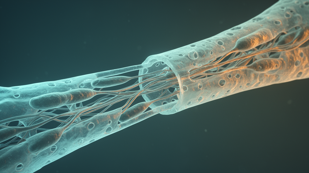
Peripheral nerve repair depends fundamentally on the dynamic plasticity of Schwann cells, which undergo profound phenotypic reprogramming following tissue trauma to clear degenerating myelin debris, secrete neurotrophic factors, construct cellular guidance tracks known as bands of Büngner, and orchestrate target reinnervation. In a systematic scientific review published in Frontiers in Cellular Neuroscience, researchers Zhiyu Li, Lanxin Luo, Jiming Xie, Jingbo Wu, Juan Xing, and Ying Wan synthesized findings from literature indexed in PubMed through October 1, 2026, evaluating how biochemical and mechanical cues within the injury microenvironment direct glial fate and how modern biomaterial platforms are engineered to modulate these signaling pathways. The authors established that following axonotmesis or severe neurotmesis, distal stump Schwann cells transition from quiescent myelinating units into specialized repair cells governed by interconnected molecular networks. However, endogenous repair Schwann cells exhibit progressive functional exhaustion over extended periods and long anatomical gaps, undergoing cellular aging and downregulating vital repair programs. Consequently, investigators are developing advanced biomaterial scaffolds, nerve guidance conduits, and responsive hydrogels designed to spatiotemporally direct glial reprogramming and enhance structural and functional recovery across peripheral nerve lesions.
At the center of Schwann cell transdifferentiation stands the basic leucine zipper transcription factor c-Jun, recognized as an indispensable master regulator that orchestrates the shift from a differentiated, myelin-maintaining phenotype toward an active repair state. Following nerve transection or crush injuries, mitochondrial calcium release mediated by voltage-dependent anion channel 1 stimulates the c-Jun N-terminal kinase cascade, triggering rapid and robust c-Jun upregulation within distal Schwann cells. Elevated c-Jun coordinates a comprehensive transcriptional repertoire, activating cytokine secretion, autophagy programs, and essential neurotrophic signals, including brain-derived neurotrophic factor and nerve growth factor, while driving the demyelination of severed distal axonal segments. Experimental genetic ablations confirm that while Schwann cell-specific c-Jun deletion does not disturb baseline embryonic development or uninjured nerve architecture, it severely impairs trophic provision, delays myelin breakdown, disrupts the structural formation of bands of Büngner, and stalls axonal regeneration. Nevertheless, c-Jun expression must decline during late repair phases to permit remyelination, as sustained elevation represses myelin-associated genes, inspiring engineered poly(lactide-co-glycolide) nanofiber architectures and erythropoietin-loaded alginate hydrogels formulated to support early c-Jun activation while permitting subsequent redifferentiation.
Working in close association with c-Jun, canonical Notch signaling serves as an essential, context-dependent modulator of glial plasticity, operating through transmembrane receptors Notch1 through Notch4 and ligands including Jagged1, Jagged2, and Delta-like molecules. Upon ligand interaction, sequential proteolytic cleavage by enzymes including gamma-secretase liberates the Notch intracellular domain, which translocates to the nucleus to assemble transcription complexes that stimulate downstream effectors such as Hes1 and Hes5. During early post-injury intervals, transient Notch activation assists dedifferentiation by directly repressing pro-myelinating transcription factors such as Krox20, lowering the barrier for entry into the regenerative state. Investigators note an asymmetrical crosstalk wherein c-Jun functions as the dominant transcriptional driver of repair genes, whereas Notch functions primarily as a permissive facilitator of cellular plasticity without exhibiting direct reciprocal inhibition. Prolonged Notch activation hinders functional remyelination, prompting researchers to develop local biomaterial delivery matrices, such as three-dimensional collagen hydrogels and functionalized nanofiber scaffolds presenting immobilized Jagged1, aiming to capture early repair benefits in preclinical models while circumventing broad systemic dysregulation.
Axon-derived communication mediated by neuregulin 1 and its corresponding ErbB receptor tyrosine kinases, predominantly ErbB2 and ErbB3 heterodimers, supplies the instructive cues necessary for Schwann cell survival, radial sorting, and eventual remyelination. Transmembrane type III neuregulin 1 acts as a strict physical molecular ruler at the axon-glia interface, dictating myelin sheath thickness through localized spatial positioning, while soluble type I isoforms govern early glial migration and proliferative survival. Ligand engagement triggers downstream signaling via mitogen-activated protein kinase and extracellular signal-regulated kinase cascades alongside the docking protein Gab1, which couples receptor activation directly to the induction of the master pro-myelinating transcription factor Krox20, also termed EGR2. In chronically denervated distal nerve stumps, prolonged loss of axonal contact causes severe neuregulin 1 downregulation and glial atrophy, substantially impairing delayed regenerative interventions. To mitigate this decline, biomaterial strategies utilize sustained-release hydrogels, conduits functionalized with extracellular vesicles, or pharmacological adjuvants such as metformin and pulsed ultrasound, all designed to reconstitute ErbB signaling, preserve Schwann cell responsiveness, and stimulate robust myelin protein synthesis during regenerative axonal re-extension.
Intracellularly, the phosphatidylinositol 3-kinase, protein kinase B, and mechanistic target of rapamycin signaling axis operates as a pivotal metabolic and survival hub downstream of neuregulin 1 and extracellular matrix integrin engagement. During early post-injury stages, moderate pathway activation preserves mitochondrial respiration, promotes metabolic reprogramming, and sustains Schwann cell viability within a nutrient-deprived, inflammatory microenvironment populated by recruited macrophages and stromal fibroblasts. However, the timeline of pathway activity remains critical, as excessive or prolonged mechanistic target of rapamycin activation downstream suppresses cellular autophagy and hinders timely myelinophagy, which is required for effective myelin debris clearance. Experimental biomaterial conduits incorporating extracellular vesicle-enriched decellularized matrices, directional microchannels, and wireless magnetoelectric hydrogels such as DG/FBD illustrate the therapeutic modulation of this cascade, upregulating repair chemokines such as CXCL12 to inhibit excessive mTOR signaling and enhance autophagic clearance. Furthermore, scaffolds functionalized with natural extracellular matrix ligands like laminin and fibronectin leverage integrin engagement to stimulate balanced Akt signaling, promoting glial adhesion, elongation, and cytoskeletal stability along the regenerating bridge.
Transforming growth factor-beta signaling introduces an additional layer of microenvironmental control, transmitting signals through type I and type II serine-threonine kinase receptors and downstream Smad2 and Smad3 transcriptional regulators, as well as non-canonical mitogen-activated protein kinase pathways. In Schwann cell cultures, transforming growth factor-beta 1 markedly accelerates cell motility and invasive migration through the upregulation of matrix metalloproteinases, specifically MMP-2 and MMP-9, which break down inhibitory debris and allow cells to mobilize toward damaged zones. Experimental knockdown of Smad2 completely abolishes this factor-induced MMP-2 expression and cellular migration, confirming the pathway’s direct role in early tissue remodeling during Wallerian degeneration. Extracellular matrix components, including matrilin-2, also interface with the transforming growth factor-beta and bone morphogenetic protein 7 signaling axes to facilitate cell movement. However, because unrestrained pathway activity can stimulate persistent fibrosis, excessive scar tissue formation, and chronic inflammatory changes, biomaterial approaches increasingly focus on degradable hydrogel formulations capable of transient, localized growth factor regulation to prevent pathological fibrotic remodeling within nerve gaps.
Complementing growth factor pathways, the Wnt and beta-catenin signaling cascade exhibits divergent, tissue-specific functions between central and peripheral nervous systems, demanding precise therapeutic calibration. In central nervous system models of demyelination, sustained Wnt activation functions as a differentiation block that represses Krox20 and prevents oligodendrocyte precursor maturation, explaining remyelination failure in multiple sclerosis. In contrast, peripheral studies demonstrate that Wnt and beta-catenin signaling actively supports Schwann cell proliferation and directional migration following injury, with Celsr2 gene silencing shown to diminish nuclear beta-catenin accumulation and impair glial motility in vitro. Pharmacological agents including small-molecule inhibitors such as XAV939, indomethacin, and celastrol illustrate that modulating specific pathway nodes can tune glial behavior, although peripheral nervous system-specific evidence remains less extensive than central nervous system literature. Engineered nerve scaffolds are consequently exploring the controlled presentation of immobilized Wnt ligands or stage-specific antagonists, aiming to stimulate initial Schwann cell expansion and migration across critical-sized nerve defects without imposing delayed obstacles to remyelination.
Schwann cell function is equally governed by physical forces through specialized mechanotransduction networks, primarily focal adhesion kinase and the Hippo-Yes-associated protein and transcriptional coactivator with PDZ-binding motif cascade. Extracellular matrix proteins, including laminin, type I collagen, and fibronectin, engage cell-surface integrins to trigger focal adhesion kinase autophosphorylation, initiating downstream cytoskeletal remodeling that enables glial spreading, radial sorting, and alignment into organized migratory columns. Simultaneously, the Hippo pathway’s core effectors, YAP and TAZ, display an atypical regulatory paradigm in peripheral glia; unlike typical epithelial tissues where nuclear YAP and TAZ stimulate proliferation and suppress differentiation, peripheral Schwann cells strictly require nuclear YAP and TAZ to activate Krox20 and drive developmental myelination and adult myelin sheath maintenance. Following nerve transection, nuclear YAP and TAZ temporarily disappear from Schwann cells and reappear only upon renewed contact with regenerating axons. Biomimetic conduits with aligned microgrooves, tunable viscoelasticity, and chiral hydrogel matrices containing right-handed L-phenylalanine fibers harness these mechanosensitive pathways to stimulate endogenous Schwann cell elongation and promote coordinated functional recovery.
Modern engineering paradigms have steadily transitioned nerve repair platforms from inert silicone or synthetic polymer tubes into intelligent, microenvironment-responsive biomaterial systems capable of actively modulating cellular signaling dynamics. Researchers evaluate advanced constructs such as electrohydrodynamic jet three-dimensional printed conduits, aligned polycaprolactone fibers functionalized with heparin-bound neurotrophic factors, brain-derived decellularized extracellular matrix shells, and chiral nanostructured hydrogels in preclinical rat sciatic nerve defect models. These active scaffolds provide crucial biomimetic topographies, controlled viscoelastic stress relaxation, and localized cargo delivery, shielding transplanted or migrating Schwann cells from destructive mechanical shear while directing their phenotypic trajectory. Nevertheless, the review emphasizes that pathway involvement across these experimental models is predominantly inferred from correlated marker changes or histological improvements rather than confirmed through cell-specific genetic ablation or pharmacological inhibition. Disentangling the independent contributions of physical alignment, matrix mechanics, and biochemical ligand presentation requires rigorous, cell-resolved mechanistic validation before therapeutic benefits observed in animal defect models can be attributed conclusively to defined Schwann cell signaling pathways.
Despite substantial preclinical progress in repairing short-gap nerve injuries, several formidable scientific and translational hurdles impede the clinical implementation of Schwann cell-modulating biomaterials. Most current scaffolding architectures provide static mechanical and chemical characteristics, whereas native peripheral nerve repair demands highly dynamic, stage-specific signaling transitions from early dedifferentiation and debris clearance to late axonal contact-dependent remyelination. Additionally, clinical translation faces shortages of autologous Schwann cell sources, prolonged denervation-induced microenvironmental hostility in long-gap human injuries, and manufacturing challenges surrounding batch reproducibility, decellularization purity, and regulatory compliance. Future therapeutic paradigms will increasingly depend on responsive, enzyme-cleavable hydrogels, dynamic-stiffness substrates that mimic basal lamina maturation, and single-cell multi-omics profiling to identify precise molecular targets and intervention windows. While complementary clinical strategies like regenerative peripheral nerve interfaces and specialized rehabilitation alleviate symptoms and facilitate prosthetic integration, translating advanced biomaterial-mediated Schwann cell reprogramming into definitive functional restoration requires comprehensive long-term evaluation in standardized large-animal models.
Subject of Research: Schwann cell reprogramming and biomaterial-mediated signaling modulation in peripheral nerve regeneration
Article Title: Modulating Schwann cell reprogramming with biomaterials: targeting signaling networks for peripheral nerve regeneration
Article References: Li, Z., Luo, L., Xie, J., Wu, J., Xing, J., & Wan, Y. (2026). Modulating Schwann cell reprogramming with biomaterials: targeting signaling networks for peripheral nerve regeneration. Frontiers in Cellular Neuroscience, 20, Article 1959013. https://doi.org/10.3389/fncel.2026.1959013
Image Credits: AI Generated
DOI: 10.3389/fncel.2026.1959013
Keywords: Schwann cells, peripheral nerve regeneration, biomaterials, nerve guidance conduits, c-Jun, Notch signaling, NRG1 ErbB, PI3K Akt mTOR, TGF-beta Smad, Wnt beta-catenin, integrin FAK, Hippo YAP TAZ, mechanotransduction, bands of Bungner, remyelination, hydrogels, Wallerian degeneration, cellular reprogramming
News Source: Gregory Coleman. (November 11, 2026). Biomaterial Platforms Target Schwann Cell Reprogramming Pathways for Peripheral Nerve Regeneration. Scienmag.