An integrated computational framework called Feature Optimization and Cascaded Unified Screening, or FOCUS, combined with a Support Vector Machine classifier, achieved an accuracy of 0.902 and an area under the receiver operating characteristic curve of 0.909 for classifying isocitrate dehydrogenase mutation status in a study cohort of 73 glioma patients.
Accurate classification of isocitrate dehydrogenase mutation status is essential for the diagnosis and clinical management of adult-type diffuse gliomas under the 2021 World Health Organization Classification. However, standard histological evaluations and molecular diagnostic workflows are labor-intensive, time-consuming, expensive, and frequently constrained by limited accessibility across clinical pathology laboratories.
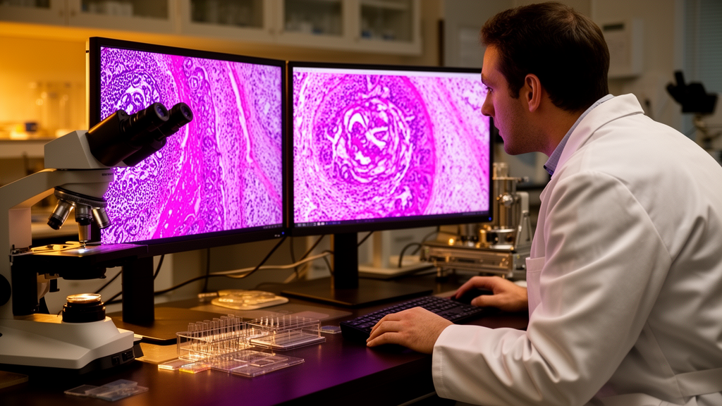
Although recent advancements in artificial intelligence have facilitated automated and objective glioma assessment directly from histopathological images, existing computational tools still encounter difficulties in capturing complex histological structures and molecular characteristics. To resolve these challenges, the FOCUS system begins by dividing digital glioma pathology slide images into smaller, standardized image patches for systematic evaluation.
From each individual patch, a Vision Transformer model extracts interpretable visual features that correspond to nuclear morphology and the expression of proteins linked to tumor progression. This localized extraction step isolates detailed cellular properties directly from the digital tissue specimens.
Following patch-level extraction, a Patch-based Graph Convolutional Network aggregates spatial contextual information across the slide to model both localized tissue patterns and broader global tumor heterogeneity. The pipeline then applies a cascaded feature selection strategy to refine biomarker identification before supplying the optimized feature subset to a machine learning classifier for final prediction.
In evaluations using the Support Vector Machine classifier, the proposed framework demonstrated the capacity to computationally distinguish isocitrate dehydrogenase-mutant tumors, including astrocytoma and oligodendroglioma, from isocitrate dehydrogenase-wildtype glioblastoma. The authors concluded that the FOCUS architecture offers an interpretable and robust computational approach for molecular subtype classification, while noting that the reported performance was established on an initial dataset of 73 patients.
Subject of Research: Machine learning classification of glioma IDH mutation status from histopathological images
Article Title: FOCUS: a feature optimization and cascaded unified screening strategy for computer-aided pathology-based glioma molecular subtype classification
Article References: Su, Y., Li, F., Yang, H., Bai, J., & Lu, J. (2026). FOCUS: a feature optimization and cascaded unified screening strategy for computer-aided pathology-based glioma molecular subtype classification. Diagnostic Pathology, 21(1), Article 84. https://doi.org/10.1186/s13000-026-01836-1
Image Credits: AI Generated
DOI: 10.1186/s13000-026-01836-1
Keywords: FOCUS framework, IDH mutation status, glioma classification, diffuse gliomas, histopathological images, Vision Transformer, PatchGCN, Support Vector Machine, tumor heterogeneity, nuclear morphology, astrocytoma, oligodendroglioma, glioblastoma, cascaded feature selection, 2021 WHO Classification, computational pathology, molecular subtyping
News Source: Ophelia Keating. (November 11, 2026). FOCUS Model Achieves 0.902 Accuracy Classifying Glioma IDH Mutation Status. Scienmag.