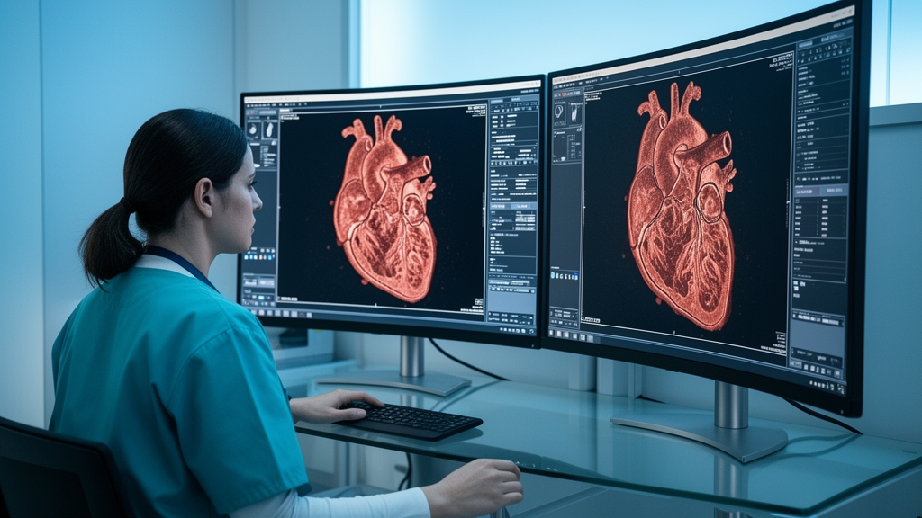
A retrospective radiological investigation conducted at the University Hospital Bonn has demonstrated that quantitative radiomics coupled with supervised machine learning can reliably detect fatal myocardial infarction on noncontrast postmortem computed tomography scans. Cardiovascular disease and acute myocardial infarction remain among the principal causes of death worldwide, responsible for roughly one-third of all mortalities in people over the age of thirty-five. While conventional forensic autopsy continues to serve as the definitive diagnostic standard for determining cause of death, forensic and medical pathologists increasingly employ whole-body postmortem computed tomography as an informative, noninvasive adjunct. By evaluating subtle, sub-visual gray-level texture patterns across the left ventricular myocardium, the research team demonstrated that computational models can differentiate between autopsy-confirmed myocardial infarction cases and non-cardiac control cases with high diagnostic accuracy.
The study protocol, approved by the local institutional ethics committee under reference number 2025-165-BO, evaluated imaging archives curated by the Institute of Forensic Medicine at University Hospital Bonn. Investigators screened 619 consecutive postmortem computed tomography examinations, applying rigorous inclusion and exclusion criteria that culminated in a well-defined cohort of 106 deceased individuals. The experimental group comprised 55 deceased subjects whose definitive cause of death was established at forensic autopsy as acute myocardial infarction or acute reinfarction. The control cohort consisted of 51 decedents who died of non-cardiac causes without evidence of cardiac pathology upon comprehensive examination. Autopsy evaluation categorized infarction into acute or subacute presentations, characterized macroscopically by pale or yellowish myocardial tissue discoloration, and acute reinfarctions, marked by fresh pale ischemic zones immediately adjacent to grayish-white fibrotic scars. Routine histological analyses using hematoxylin and eosin staining verified the microscopic hallmarks of infarction, displaying characteristic wavy, hypereosinophilic myocytes, cytoplasmic cross-striation loss, karyolysis, and dense neutrophilic infiltrates in acute lesions, alongside collagenous scar formations in recurring infarction cases.
Computed tomography acquisitions were performed on a dedicated sixteen-slice multi-detector scanner, specifically the SOMATOM Emotion 16 manufactured by Siemens Healthineers in Erlangen, Germany. Standardized volumetric scanning protocols operated at a tube voltage of 130 kilovolts and an exposure tube current of 100 milliamperes, generating soft-tissue reconstructions through a B30s convolution kernel. Scans utilized a slice thickness of 1.5 millimeters, a reconstruction increment of 1.0 millimeter, a spiral pitch of 1.5, and a broad field of view measuring 500 millimeters. Transverse image slices were reconstructed on a matrix size of 512 by 512 pixels with an isotropic in-plane pixel spacing of 0.97 by 0.97 millimeters. A unique physical phenomenon inherent to uncirculated postmortem blood, known as postmortem sedimentation, aided visualization without artificial contrast administration. In the absence of cardiac output and systemic circulation, intravascular blood separates gravitationally into a hypodense clear serum supernatant and a hyperdense dependent sediment of cellular erythrocytes, creating a distinct natural density contrast that cleanly delineates the endocardial border from intracavitary contents.
Segmentation of the entire anatomical left ventricular myocardium was carried out in consensus by two experienced radiological readers, possessing over ten years and over two years of professional expertise, respectively. Working within the open-source software platform 3D Slicer, the readers employed the Grow from Seeds tool in the Segment Editor module, which executes an enhanced GrowCut algorithm. Seed regions manually defined across axial, coronal, and sagittal planes were automatically expanded, followed by manual correction to exclude non-myocardial tissue. A joint smoothing boundary refinement algorithm using a smoothing factor of 0.5 was systematically applied, generating 212 separate left ventricular volumes of interest across the cohort. Inter-observer reproducibility between the readers was quantitatively verified through two-way mixed-effects intraclass correlation coefficient analysis with absolute agreement. Feature extraction complied with guidelines established by the Image Biomarker Standardization Initiative using the SlicerRadiomics interface powered by PyRadiomics library version 3.1.0. All volumetric datasets underwent spatial resampling to an isotropic voxel grid of one cubic millimeter using linear interpolation.
The computational feature-extraction pipeline generated 1,037 mathematical radiomic features per case, designed to capture subtle textural architecture and structural heterogeneity across diverse spatial domains. This feature repository encompassed 107 original raw features, comprising 18 first-order histogram metrics, 14 three-dimensional shape characteristics, and 75 higher-order textural matrix indices. Beyond unfiltered data, the framework applied spatial Laplacian-of-Gaussian spatial bandpass filters at scaling parameters of sigma 3.0 millimeters and sigma 5.0 millimeters to accentuate fine and coarse edge transitions, alongside comprehensive three-dimensional discrete wavelet transformations combining directional low-pass and high-pass spatial filtering. Prior to analytical modeling, all extracted numerical values underwent sign-based logarithmic mathematical transformations to normalize extreme variance distributions while preserving negative polarity. Reproducibility filtering subsequently excluded unstable features susceptible to manual segmentation variations, retaining 586 robust radiomic features demonstrating an intraclass correlation coefficient of 0.75 or higher. Redundancy was mitigated by calculating pairwise Spearman correlation coefficients within each training fold, systematically dropping redundant variables when correlation coefficients exceeded 0.8 within related feature classes.
Model training and evaluation adhered to a nested stratified cross-validation architecture consisting of five outer folds and five inner cross-validation folds to prevent information leakage and ensure unbiased performance reporting. The outer validation folds remained strictly quarantined as held-out test sets, while inner validation folds handled hyperparameter tuning via systematic grid searching, correlation pruning, and feature selection. Investigators evaluated three distinct feature-handling paradigms: a baseline strategy incorporating all reproducibility-filtered features without additional selection, an embedded random forest ranking method using mean decrease in impurity, and a wrapper-based recursive feature elimination algorithm using logistic regression. Seven supervised machine-learning algorithms implemented in Python using scikit-learn, XGBoost, and CatBoost were evaluated, including logistic regression, support vector machines, k-nearest neighbors, naïve Bayes, random forest, XGBoost, and CatBoost. Model performance across the held-out outer folds was quantitatively measured using the area under the receiver operating characteristic curve, alongside overall classification accuracy, balanced accuracy, precision, sensitivity, specificity, and F1-scores.
Across all evaluated combinations of machine-learning algorithms and dimensionality reduction strategies, regularized logistic regression operating without additional feature selection achieved the highest and most balanced diagnostic performance. This top-performing model recorded a mean area under the receiver operating characteristic curve of 0.817 with a standard deviation of 0.12 across outer folds. The model demonstrated an overall classification accuracy of 0.746 plus or minus 0.05, a clinical sensitivity of 0.764 plus or minus 0.14, and a diagnostic specificity of 0.724 plus or minus 0.13. Support vector machines and random forest classifiers without additional selection exhibited highly comparable discriminative capability, delivering mean area under the curve metrics of 0.814 plus or minus 0.10 and 0.814 plus or minus 0.08, respectively. The support vector machine yielded a diagnostic accuracy of 0.745 plus or minus 0.05 with a sensitivity of 0.782 plus or minus 0.10 and specificity of 0.704 plus or minus 0.16, while random forest achieved an accuracy of 0.707 plus or minus 0.04, sensitivity of 0.691 plus or minus 0.05, and specificity of 0.726 plus or minus 0.11.
The remaining supervised classifiers generally yielded mean area under the curve estimates spanning between 0.700 plus or minus 0.03 and 0.783 plus or minus 0.12, with only naïve Bayes without selection and extreme gradient boosting under recursive feature elimination dropping below the 0.70 threshold. When comparing dimensionality reduction methodologies, the baseline approach without additional selection outperformed random forest selection and recursive feature elimination across most algorithmic architectures, including logistic regression, support vector machines, k-nearest neighbors, and random forests. Notably, stability tracking identified four radiomic features that were independently prioritized by both random forest ranking and recursive feature elimination in two or more outer cross-validation folds. These consistently selected biomarkers included the 90th percentile intensity from first-order metrics derived from Laplacian-of-Gaussian filtering at sigma 5.0 millimeters, Strength calculated from the neighborhood gray-tone difference matrix at sigma 5.0 millimeters, and two directional gray level co-occurrence matrix features: Informational Measure of Correlation 1 under wavelet low-high-high decomposition, and Correlation under wavelet high-high-high spatial decomposition.
Because postmortem computed tomography captures tissue in a static state, the diagnostic capacity of these models is entirely independent of dynamic blood flow, active myocardial contraction, or exogenous iodinated contrast distribution. The authors observed that relatively straightforward linear and boundary-based models, such as logistic regression and support vector machines, matched or outperformed complex gradient-boosted decision trees.
The findings offer practical implications for forensic pathology and medicolegal death investigations where formal internal autopsies are impossible or restricted. Conventional postmortem examinations are frequently precluded by severe shortages of certified forensic pathologists, institutional resource constraints, legal limitations, or objections raised by families on religious, cultural, or personal grounds, notably within Muslim and Jewish traditions. Non-invasive approaches are likewise often preferred in perinatal death investigations, where imaging like postmortem computed tomography may represent the only way to determine or narrow down the cause of death. Furthermore, eliminating the need for complex, costly postmortem computed tomography angiography saves substantial procedural time and institutional expenditure. Nonetheless, the investigators emphasized several methodological boundaries, noting that this exploratory single-center study analyzed a retrospective cohort of 106 cases scanned on a single machine. The research team noted that broader multicenter investigations across variable postmortem intervals, diverse scanner vendors, and varied reconstruction kernels are necessary to confirm generalizability, alongside prospective clinical studies exploring whether unenhanced radiomic analysis can eventually assist living patients who cannot receive iodinated contrast agents.
Subject of Research: Machine learning and radiomics analysis of noncontrast postmortem computed tomography for myocardial infarction detection
Article Title: Detection of myocardial infarction by postmortem CT combined with radiomics-based machine-learning analysis
Article References: Barman, N. C., Rastogi, A., Radbruch, A., Luetkens, J. A., Wittschieber, D., & Hahnemann, M. (2026). Detection of myocardial infarction by postmortem CT combined with radiomics-based machine-learning analysis. European Radiology Experimental, 10(1), Article 130. https://doi.org/10.1186/s41747-026-00800-4
Image Credits: AI Generated
DOI: 10.1186/s41747-026-00800-4
Keywords: Postmortem computed tomography, Myocardial infarction, Radiomics, Machine learning, Forensic pathology, Logistic regression, Support vector machine, Random forest, Image texture analysis, Cardiac imaging, Feature extraction, Autopsy, Left ventricle, Tissue characterization, Noncontrast CT, PyRadiomics, Stratified cross-validation
News Source: Teresa Odom. (November 11, 2026). Machine Learning and Postmortem CT Radiomics Accurately Identify Fatal Myocardial Infarction. Scienmag.