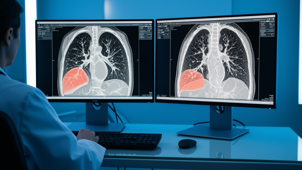
Researchers Mahender Erukala and Suresh Kumar Sanampudi have introduced a new deep learning framework designed to improve the segmentation of lung nodules in computed tomography images. The model, named CLAHE-RATSNet, addresses specific technical challenges that often hinder the accurate delineation of these nodules. Published in BMC Medical Imaging, the study presents a click-guided segmentation network that utilizes residual attention for a two-stage processing approach. The primary goal of this framework is to enhance computer-aided diagnosis, treatment planning, and the early detection of lung cancer by overcoming common imaging obstacles.
The segmentation of lung nodules is a difficult task in clinical practice due to several inherent factors. These include low contrast in the images, the small physical size of the nodules, and weak boundaries that make them hard to distinguish. Additionally, nodules often resemble surrounding vessels and lung tissues, which complicates the identification process. To solve these problems, the authors developed a system that combines image enhancement techniques with advanced neural network architectures to provide more precise localization and boundary correction.
The proposed framework integrates Contrast Limited Adaptive Histogram Equalization, or CLAHE, with a Gaussian click map. This click map is generated from the centroid of the nodules to provide spatial guidance, while CLAHE provides enhanced CT contrast at the local level.
The architecture of CLAHE-RATSNet employs a two-stage segmentation process. A first-stage attention network is utilized for the coarse localization of the nodules. Following this initial step, a second-stage residual refinement module is applied to correct boundary errors.
Experiments were conducted using the LIDC-IDRI computed tomography images to evaluate the performance of the new framework. The results of CLAHE-RATSNet were compared against several baseline models to determine its relative effectiveness.
Under an oracle-guided test condition, the proposed model achieved a Dice score of 0.9238 and an Intersection over Union, or IoU, of 0.8610. The model also recorded a precision of 0.9239 and a recall of 0.9238.
Statistical analyses were performed to compare CLAHE-RATSNet with the evaluated baseline models. Patient-level paired analyses showed statistically significant differences in Dice, IoU, precision, and recall. These differences remained significant after applying the Holm correction for multiple comparisons.
It is important to note that these findings characterize segmentation performance under a controlled oracle-guided evaluation protocol. The results should not be interpreted as end-to-end lesion-localization or fully automatic clinical performance.
As the study is published early to provide faster access to peer-reviewed research, it remains subject to further edits before the final version of record is released.
Subject of Research: Deep learning framework for lung nodule segmentation in CT images
Article Title: CLAHE-RATSNet: a deep learning framework for accurate lung nodule segmentation in computed tomography images
Article References: Erukala, M., & Sanampudi, S. K. (2026). CLAHE-RATSNet: a deep learning framework for accurate lung nodule segmentation in computed tomography images. BMC Medical Imaging. https://doi.org/10.1186/s12880-026-02991-z
Image Credits: AI Generated
DOI: 10.1186/s12880-026-02991-z
Keywords: lung nodule segmentation, computed tomography, deep learning, medical imaging, CLAHE-RATSNet, computer-aided diagnosis, image enhancement, attention network, deep, learning, framework, accurate
News Source: Ophelia Keating. (November 10, 2026). New Deep Learning Model Segments Lung Nodules in CT Scans. Scienmag.