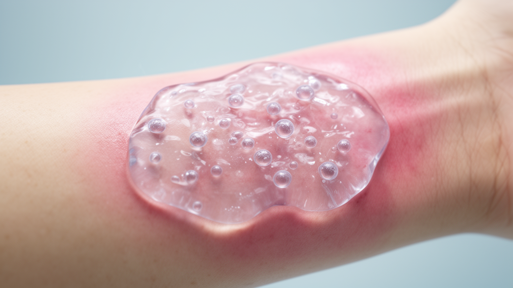
Researchers have developed a multifunctional bio-derived hydrogel system that integrates adipose-derived stem cells (ADSCs) to promote healing in diabetic foot ulcers. Published in Materials Today Bio, the study demonstrates that this composite scaffold, composed of gelatin methacryloyl (GelMA), glycyrrhizin (GA), and iron ions (Fe2+), effectively addresses the complex pathological features of chronic diabetic wounds. The system is designed to simultaneously modulate the hostile wound microenvironment and support tissue regeneration, offering a potential advancement over current standard treatments that often fail to resolve multifactorial cellular dysfunctions.
Diabetic foot ulcers (DFUs) are a serious complication of diabetes mellitus, affecting an estimated 15-25% of individuals with the disease over their lifetime. The pathogenesis of these wounds is characterized by a stalled healing process involving persistent hyperglycemia, chronic inflammation, impaired angiogenesis, and susceptibility to bacterial infection. Unlike acute wounds, the diabetic wound microenvironment is hostile, marked by excessive reactive oxygen species (ROS) and a dysfunctional extracellular matrix (ECM) that fails to support cellular migration and tissue repair. Current standard treatments, such as debridement and revascularization, frequently fail to address these underlying cellular dysfunctions, creating an urgent need for advanced regenerative therapies.
The study focuses on the therapeutic potential of ADSCs, which can regulate inflammatory responses, stimulate new blood vessel formation, and support epithelialization through the paracrine secretion of bioactive mediators. However, the unfavorable microenvironment of diabetic wounds substantially limits the clinical effectiveness of ADSC therapy. High levels of oxidative stress and inflammatory cytokines lead to rapid stem cell apoptosis and poor retention at the injury site. Furthermore, direct injection of cells often results in leakage and a lack of sustained paracrine signaling. Existing hydrogel carriers often lack sufficient bioactivity or fail to provide rapid ROS scavenging kinetics to protect the encapsulated payload, while some natural hydrogels suffer from rapid degradation or insufficient mechanical support.
To address these challenges, the researchers engineered a dual-network hydrogel system. The GelMA component establishes a covalent network that mimics the mechanical stiffness of the native ECM, creating a matrix that supports cell attachment and migration. The incorporation of GA, a natural triterpenoid saponin, imparts potent anti-inflammatory and antioxidant capabilities essential for neutralizing the oxidative storm characteristic of diabetic wounds. Crucially, the addition of Fe2+ ions serves as a functional bridge, forming stable coordinate bonds with GA to reinforce the hydrogel’s structural integrity through a supramolecular secondary network. This coordination also exhibits a powerful synergistic antibacterial effect with GA, addressing the high bacterial burden typical of chronic diabetic ulcers.
Characterization of the hydrogel revealed a porous architecture suitable for cell encapsulation. Scanning electron microscopy showed a structure that facilitated the uniform distribution of ADSCs within the matrix. Rheological properties indicated that the GelMA/GA/Fe hydrogel exhibited the highest storage modulus, suggesting enhanced mechanical stiffness resulting from the integration of the GA-Fe coordination network with the photocrosslinked GelMA network. The swelling behavior of the composite hydrogel remained relatively stable at approximately 294%, compared to 359% for GelMA alone, indicating that the dual-network structure restricts excessive water uptake while maintaining hydrogel integrity. Release studies using fluorescein isothiocyanate-labeled bovine serum albumin as a model protein showed that the composite hydrogel provided slower, more controlled release, supporting its potential as a scaffold for localized delivery.
In vitro biocompatibility assays confirmed that the hydrogel extracts did not impair cellular metabolic activity. ADSCs exposed to hydrogel extracts exhibited higher relative cell viability than untreated controls at both 24 and 48 hours. Hemolysis assays showed that all hydrogel extract groups exhibited hemolysis rates below approximately 5%, suggesting good hemocompatibility under the tested conditions. Iron-related safety evaluations indicated that the GelMA network may regulate Fe2+ release and reduce local iron-associated deposition, as evidenced by lower Prussian blue positive staining areas compared to the GA/Fe group. Histological analysis of major organs in animal models showed that tissue architecture remained largely intact across the experimental groups.
The effects of the hydrogel on wound closure-related cellular responses were evaluated using scratch assays under high-glucose conditions. The GelMA/GA/Fe@ADSCs group exhibited the highest scratch closure rate in L929 cells after 24 hours, reaching approximately 68%, compared with approximately 14% in the control group. A similar trend was observed in HaCaT keratinocytes, with the composite group achieving approximately 85% scratch closure. These findings suggest that combining the bioactive dual-network hydrogel with ADSCs enhances cellular responses associated with wound closure. In endothelial network formation assays, the GelMA/GA/Fe@ADSCs group showed the highest mesh count, indicating significant improvement in capillary-like network formation compared to the cell-free hydrogel.
Western blotting analysis revealed that the ADSC-loaded hydrogel increased the phosphorylation of AKT and mTOR, alongside higher expression of HIF-1α and VEGF. These changes were attenuated by pretreatment with LY294002, a PI3K inhibitor, indicating that the molecular response was sensitive to PI3K/AKT/mTOR-associated signaling. This suggests that these pathways may contribute to the pro-angiogenic activity of the composite system. Macrophage phenotype changes were evaluated by flow cytometry, showing that the GelMA/GA/Fe@ADSCs group shifted the macrophage phenotype toward a higher CD206/CD86 ratio. This indicates a polarization tendency toward an M2-like phenotype, suggesting that ADSCs contribute to the immunomodulatory effects of the composite system by promoting a repair-supportive inflammatory environment.
The therapeutic efficacy of the system was evaluated in a diabetic wound model using rats rendered diabetic by intraperitoneal administration of streptozotocin. Wound areas were monitored over 11 days, and the GelMA/GA/Fe@ADSCs group showed the smallest residual wound area, approaching complete closure by day 11. Histological evaluation by H&E and Masson’s trichrome staining demonstrated that the ADSC-loaded group exhibited the smallest measured wound width and more extensive collagen deposition. The quantified collagen fraction in this group reached approximately 58%, compared with 47% for the cell-free GelMA/GA/Fe group and 10% for the control group. These results support the therapeutic benefit of combining the dual-network hydrogel with ADSCs in diabetic wound repair.
In an infected wound model, the cell-free GelMA/GA/Fe hydrogel also showed significant therapeutic performance, approaching complete closure by day 12. Local bacterial burden was quantified by colony counting of wound tissue samples, revealing that GA/Fe-containing hydrogels reduced the Staphylococcus aureus burden. The GelMA/GA/Fe group showed the lowest bacterial burden, although the persistence of recoverable colonies indicates bacterial reduction rather than complete eradication. Immunohistochemical and fluorescence staining in diabetic wound tissues revealed more abundant vascular structures in the GelMA/GA/Fe@ADSCs group, accompanied by a significantly higher blood vessel density. This observation complements the enhanced endothelial network formation observed in vitro, supporting the pro-angiogenic potential of the ADSC-loaded hydrogel in the wound environment.
The improved vascularization was accompanied by changes in inflammatory markers. Immunohistochemical analysis showed higher IL-10 and lower TNF-α staining levels in the GelMA/GA/Fe@ADSCs group than in the cell-free group. CD206/iNOS immunofluorescence further revealed stronger CD206-associated signals and reduced iNOS-associated signals following treatment with the ADSC-loaded hydrogel. This marker profile is consistent with an M2-like phenotypic shift, suggesting a more repair-supportive inflammatory environment. ROS-sensitive fluorescence staining provided additional evidence of changes in the local oxidative environment, with the GelMA/GA/Fe@ADSCs group exhibiting the lowest ROS-associated fluorescence intensity. Together, the increased vascular density, altered inflammatory marker profile, and reduced ROS-associated fluorescence support complementary effects of the combined system on the wound microenvironment.
While the study demonstrates the potential of the GelMA/GA/Fe@ADSCs system to promote tissue repair, certain limitations remain. The performance of this hydrogel relative to commercially available hydrogels or conventional wound care treatments has not yet been assessed. Although the present findings support its potential to promote tissue repair, its comparative efficacy and clinical applicability require further investigation. Moreover, despite the observed short-term therapeutic benefits, long-term ADSC survival and sustained therapeutic effects in vivo remain to be established. Future studies may explore strategies such as ADSC preconditioning or genetic modification to improve the durability and efficacy of this hydrogel-based therapy. The current findings provide a foundation for understanding how multifunctional bio-derived hydrogels can address the multifactorial challenges of diabetic wound healing.
Subject of Research: Multifunctional bio-derived hydrogel loaded with ADSCs for promoting diabetic wound healing
Article Title: Multifunctional bio-derived hydrogel loaded with ADSCs for promoting diabetic wound healing
Article References: Yuan, X., Zou, H., Li, J., Chen, J., Wang, F., Zhang, J., Zhang, Q., Chen, M., Lin, Y., Zheng, D., & Chen, G. (2026). Multifunctional bio-derived hydrogel loaded with ADSCs for promoting diabetic wound healing. Materials Today Bio, 41, Article 103803. https://doi.org/10.1016/j.mtbio.2026.103803
Image Credits: AI Generated
DOI: 10.1016/j.mtbio.2026.103803
Keywords: Multifunctional, bio-derived, hydrogel, loaded, ADSCs, promoting, diabetic, wound, healing, scientific research
News Source: Gregory Coleman. (November 8, 2026). Bio-derived hydrogel with stem cells accelerates diabetic wound healing. Scienmag.