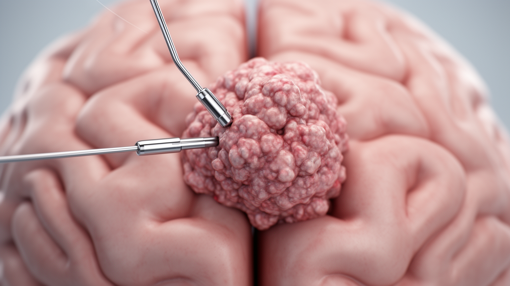
A 64-year-old man developed glioblastoma, the most common malignant primary brain tumor, two years after receiving bilateral subthalamic nucleus deep brain stimulation for Parkinson’s disease. This case report, published in the Egyptian Journal of Neurosurgery, documents the rare coexistence of these conditions and the clinical challenges associated with monitoring patients who have implanted neuromodulation hardware. The authors conducted a literature review using PubMed and Google Scholar to contextualize this specific case within the broader medical record. Their review found that only seven similar cases had been previously reported in the literature, highlighting the extreme rarity of this clinical presentation.
In this specific instance, the patient underwent subtotal resection of the tumor followed by adjuvant chemoradiotherapy. Despite these interventions, the tumor recurred, and the patient died six months later due to disease progression. The authors note that while the patient’s family history included significant malignancy, the role of such predisposing factors in this specific context remains unknown.
The study notes that many complications have been reported following deep brain stimulation procedures, including meningitis, superficial wound infection, lead exposure, intracranial hemorrhage, and neuropsychiatric disorders. In contrast, environmental risk factors such as occupational exposure to electric and magnetic fields have been proposed in the etiopathogenesis of glioblastoma. Currently, there is no consensus regarding a causal relationship between deep brain stimulation and the formation of brain tumors. While the occurrence of tumors along electrode trajectories may suggest a potential association, this phenomenon has been extremely rarely reported despite an estimated 300,000 deep brain stimulation procedures performed worldwide. The authors emphasize that the rarity of these events makes it difficult to draw definitive conclusions about causality.
Several theoretical mechanisms have been proposed to explain a potential link between chronic electrical stimulation and tumor development. These include direct effects of stimulation on cellular proliferation and apoptosis, chronic neuroinflammation and gliosis induced by implanted electrodes, localized blood–brain barrier disruption, and modulation of cell migration and growth by electric fields. Experimental studies have demonstrated that electrical stimulation may influence anti-apoptotic signaling pathways; however, the clinical significance of these findings remains unclear. Given the limited evidence and the rarity of reported cases, current data are insufficient to support a causal association between deep brain stimulation and intracranial tumorigenesis. Further studies are required to determine whether a true association exists or whether the reported cases represent coincidental findings.
The authors point out that due to the requirement for neuroimaging in deep brain stimulation targeting, all reported gliomas were identified after a variable interval following surgery. The reasons for this variability remain unclear, as does the apparent predominance of patients with tremor undergoing thalamic stimulation. It has also been hypothesized that continuous electrical stimulation may suppress tumor extension toward electrode contact points, a theory that requires further investigation.
Managing patients with both deep brain stimulation hardware and intracranial tumors presents significant logistical and clinical challenges. MRI evaluation in individuals with implanted systems is not always straightforward, particularly in centers with limited experience. However, with modern MRI-conditional deep brain stimulation systems and adherence to specific safety protocols, imaging has become more feasible. Nevertheless, variability between manufacturers and restrictive institutional protocols continue to pose practical limitations. Patients presenting with suspected intracranial tumors should be evaluated in close collaboration with radiologists to ensure accurate diagnosis and safe imaging procedures.
The case underscores the importance of a multidisciplinary approach involving neurologists, neurosurgeons, and oncologists. A low threshold for neuroimaging is recommended in deep brain stimulation patients presenting with new or atypical neurological deficits, such as hemiparesis, seizures, or disproportionate cognitive decline. Management should address hardware considerations, treatment-related risks, and the continuation of stimulation. Shared decision-making is essential, while prognosis is primarily determined by tumor pathology, particularly in high-grade gliomas. The authors conclude that further studies are needed to clarify any association between chronic electrical stimulation and tumor development, as current data are insufficient to establish a definitive link.
Altogether, only eight cases have been reported to date, including the current one, while the annual global age-standardized incidence of primary malignant brain tumors is approximately 3.7 per 100,000 in males and 2.6 per 100,000 in females. This comparison highlights the exceptionally low frequency of glioblastoma developing in the context of deep brain stimulation. The authors recommend that new atypical neurological deficits in deep brain stimulation patients should prompt neuroimaging to ensure early detection and appropriate management.
Subject of Research: Case report and literature review on glioblastoma development following deep brain stimulation
Article Title: Glioblastoma after deep brain stimulation: a case report and literature review
Article References: Parvaresh, M., Sisakht, A. M., Rohani, M., Fasano, A., & Habibi, S. A. H. (2026). Glioblastoma after deep brain stimulation: a case report and literature review. Egyptian Journal of Neurosurgery, 41(1), Article 157. https://doi.org/10.1186/s41984-026-00659-y
Image Credits: AI Generated
DOI: 10.1186/s41984-026-00659-y
Keywords: glioblastoma, deep brain stimulation, Parkinson's disease, neuro-oncology, case report, brain tumor, neuromodulation, clinical management, deep, brain, stimulation, case
News Source: Cassandra Pierce. (November 8, 2026). Glioblastoma Detected After Deep Brain Stimulation. Scienmag.