Spatial multi-omic technologies allow researchers to measure multiple layers of molecular information, including gene expression, epigenetic accessibility, and protein abundance, directly within intact tissue architectures. Despite these technological advances, modern spatial computational biology faces two major structural hurdles: vertical integration, which requires combining distinct molecular layers measured across identical spatial coordinates into a unified latent space, and horizontal integration, which requires aligning cell identities and transferring functional annotations across different experimental cohorts, tissue samples, or profiling platforms. Addressing both tasks simultaneously has historically presented severe mathematical difficulties because technical batch effects, cross-platform technical discrepancies, and measurement modalities introduce substantial domain shifts. To overcome these limitations, a research team led by Chang Xu, Shibo Liu, Yang Heng, Boxiang Liu, and colleagues introduced a geometric deep learning framework termed domain invariant representation through adaptive calibration, or DIRAC. Reporting in Nature Methods, the authors established a generalized domain adaptation architecture designed to eliminate platform-specific and modality-specific distribution shifts while strictly preserving biologically meaningful cellular identities and fine-grained spatial tissue microenvironments.
The fundamental design of DIRAC unifies vertical and horizontal data processing by framing spatial data integration through the principles of domain adaptation theory. In complex experimental settings, molecular measurements shift systematically across experimental batches, technological platforms, and omic measurement layers, yet the underlying biological cell types and functional states remain fundamentally conserved. Domain adaptation theory posits that an effective integration model must extract features that remain highly discriminative for downstream biological classification tasks while remaining completely indiscriminate with respect to domain-level variations. To implement this objective, DIRAC couples graph neural networks with an adversarial domain discriminator network. By constructing spatial and molecular graphs where cellular profiles form node attributes and physical distances define graph edges, DIRAC aggregates neighborhood contexts across intact tissues. During training, a gradient reversal layer inverts the backpropagation gradient of the domain classification loss, compelling the graph encoder to purge modality-identifying and batch-identifying technical signatures while retaining cell-state signals. The framework scales to datasets comprising millions of cells by decomposing complete cellular graphs into compact, densely connected subgraphs via a dedicated graph sampler, thereby bypassing computational bottlenecks.
For vertical multi-omic integration, DIRAC converts each measured omic layer into an independent graph by calculating spatial Euclidean distances to generate a K-nearest-neighbor spatial graph and pairing it with a molecular feature similarity graph. The intersection of these two graphs establishes a sparse consensus graph containing only cell-to-cell connections supported simultaneously by spatial proximity and molecular resemblance. Dedicated graph encoders process each individual modality to produce latent representations matching in dimensional size. The multi-omic representations are subsequently fused into a single joint latent embedding guided by an objective function combining four distinct mathematical components: domain-adversarial loss, maximum mean discrepancy to minimize divergence between omic latent distributions, multimodal consistency loss to enforce correlation between corresponding latent dimensions, and mean squared error reconstruction loss. In horizontal integration, DIRAC aligns a labeled reference dataset, designated as the source domain, with an unlabeled spatial query dataset, designated as the target domain. To prevent catastrophic distribution skewing caused by differing cell-type compositions across studies, DIRAC incorporates a pseudo-label alignment mechanism that dynamically rescales predicted class probabilities in the target domain using source-to-target expectation ratios, supported by a maximum entropy regularization term to avoid trivial single-class collapse.
To evaluate the model rigorously, the investigators first tested DIRAC on synthetic benchmarks generated by two spatial simulators: nonnegative spatial factorization, which simulates structured spatial tissues, and scMultiSim, which models diffuse and non-clustered cell distributions. On trimodal simulated datasets comprising two thousand genes, four thousand chromatin accessibility peaks, and one hundred antibody-derived tag proteins across four distinct spatial domains, DIRAC resolved clean spatial boundaries with fewer misassigned cells than competing baselines such as SpatialGLUE, MISO, and MultiVI. Quantitatively, bimodal integration with DIRAC achieved an adjusted Rand index of 0.956 and an omics mixing score of 0.748 across ten independent simulation runs, significantly outperforming the next-best evaluated methods. When tested on a real-world whole-embryo DBiT-seq dataset from mouse embryonic day 10, measuring whole-transcriptome RNA expression and twenty-two proteins at fifty-micrometer resolution, DIRAC significantly outperformed existing integration methods in both omics mixing and biological conservation metrics across ten random seeds. Ablation experiments and architectural comparisons confirmed that the consensus graph structure and domain-adversarial regularization were essential for balancing the complete mixing of disparate molecular layers with the faithful retention of delicate anatomical divisions.
Moving from technical benchmarks to complex human clinical oncology, the researchers applied DIRAC to a 10x Visium Spatial Gene and Protein Expression dataset of human glioblastoma tissue sections counterstained with DAPI and proliferating cell nuclear antigen immunofluorescence. Glioblastoma is recognized for profound cellular heterogeneity and microenvironmental niche complexity. Benchmarked against MultiVI, MISO, MaxFuse, and SpatialGLUE, DIRAC domains exhibited the highest spatial autocorrelation, achieving a Moran’s I value of 0.885. Using the Ivy Glioblastoma Atlas Project regional taxonomy, the authors resolved nine distinct spatial niches, including leading-edge white matter, extracellular matrix remodeling zones, neuron-enriched infiltration zones, tumor-associated macrophage cellular tumor cores, and a specialized microvascular proliferation niche designated as C1, corresponding to a proliferation-vascular interface. The C1 niche boundaries precisely mirrored the transition zones between proliferating tumor cells and normal brain parenchyma revealed by immunofluorescence microscopy. Deconvolution using an independent reference atlas confirmed that the C1 niche was significantly enriched for endothelial and mural cell signatures. Subsequent cell-cell communication analysis via CellChat demonstrated that the proliferation-vascular interface was driven by bidirectional signaling circuits, notably apelin signaling, wherein APLN ligands expressed in adjacent core tumor domains engaged APLNR receptors on microvascular endothelial cells along the niche boundary.
The researchers also evaluated DIRAC on horizontal label transfer tasks across severe technical dropouts and real human neuropathology cohorts. In progressive data-masking benchmarks where thirty percent of RNA and ATAC entries were randomly zeroed out, single-modality DIRAC retained an accuracy of 0.881, whereas vertically integrated bimodal DIRAC achieved an accuracy of 0.991, demonstrating that multi-omic integration confers resilience against experimental dropout. Furthermore, when specific cell types were omitted from reference datasets, DIRAC leveraged its predictive confidence thresholds to flag unassigned cells correctly, preventing forced misclassification of unseen lineages. In clinical tissue applications, the team investigated spatial transcriptomic datasets from the human dorsolateral prefrontal cortex across twelve tissue sections, where DIRAC delineated cortical layers and white matter with an accuracy of 0.839 and an area under the curve of 0.9863, outperforming support vector machines, AdaBoost, scmap, and scANVI. When applied to Stereo-seq spatial transcriptomic arrays from human prefrontal cortex Brodmann area 10 across control, moderate, and severe Alzheimer’s disease cohorts, DIRAC resolved a longstanding debate regarding cortical loss. While previous unsupervised clustering methods indicated near-total laminar collapse in advanced Alzheimer’s disease, DIRAC revealed that all six cortical laminae remained intact though markedly thinned and structurally rugged, an outcome independently corroborated by neuropathological review of matched hematoxylin and eosin tissue sections.
To establish how DIRAC performs across early mammalian developmental biology, the authors analyzed mouse embryonic stages using published single-cell and spatial datasets. Utilizing an embryonic day 8.5 whole-mouse gastrulation single-cell RNA sequencing reference, DIRAC mapped spatial cell identities across seqFISH scans of mouse embryos spanning embryonic days 8.5 to 8.75. The model accurately localized endodermal, mesodermal, and ectodermal structures, correctly distinguishing compact organ systems like the gut tube and developing heart from widely dispersed endothelial populations. When applied to the embryonic mouse forebrain, where original subclustering had merged developmental compartments into broadly overlapping prosencephalon classifications, DIRAC cleanly resolved anatomically distinct telencephalon, diencephalon, and hypothalamus zones. These predicted spatial domains were corroborated by the localized expression of canonical regional markers, including Fgf15 in the telencephalon, Wnt5a in the diencephalon, and Shh in the hypothalamus. Similarly, in a whole-embryo Stereo-seq dataset at embryonic day 16.5, DIRAC recovered finely structured anatomical features that earlier unsupervised analyses had overlooked, including developing lung airways, specialized intestinal segments, and concentrated thymic T cell populations, demonstrating consistent generalizability across embryonic stages and diverse profiling platforms.
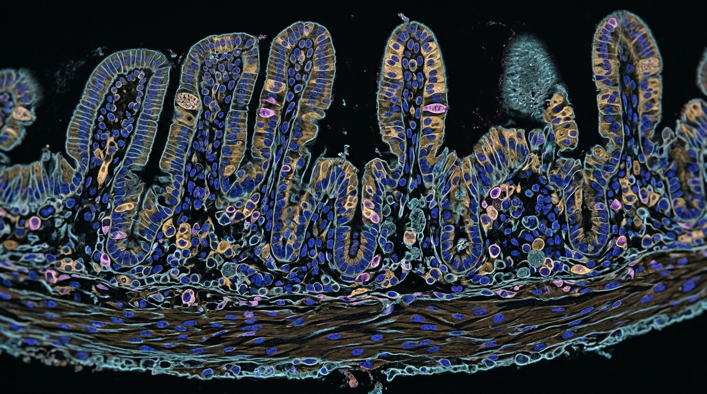
To explore lymphoid organ biology directly, the researchers generated two primary subcellular-resolution Stereo-CITE-seq datasets, capturing both whole-transcriptome mRNA and panel-specific surface proteins from adult mouse lymphoid organs. In the mouse thymus, measuring 128 surface proteins and the whole transcriptome across 173,649 cells at 500-nanometer resolution, vertical integration by DIRAC segmented the tissue into four primary histological domains: the outer capsule, the cortex, the corticomedullary junction, and the central medulla. Each region displayed distinct molecular programs, including collagen expression in the capsule, T cell antigen receptor rearrangement signatures in the cortex, antigen-presenting cell features in the junction, and immunoregulatory factors in the medulla. Increasing spatial resolution to fifty-bin increments subdivided the organ into ten distinct micro-domains following the Human Cell Atlas Common Coordinate Framework. Horizontal transfer from a single-cell CITE-seq reference comprising 71,365 annotated cells demonstrated the necessity of simultaneous multi-omic integration. While single-omic label transfers mislocalized immature thymocytes to the medullary core, DIRAC accurately confined early double-negative and double-positive T cells to the cortex while tracking their inward maturation toward mature single-positive CD4 and CD8 T cells within the medulla.
The team extended their Stereo-CITE-seq profiling to the adult mouse spleen, generating paired histological and molecular maps across 123,418 high-quality segmented single cells. Vertical multi-omic integration effectively resolved the classical architecture of the spleen, identifying the splenic capsule, the erythrocyte-rich red pulp, the white pulp, and the marginal zone, which are difficult to distinguish cleanly through hematoxylin and eosin staining alone. By integrating a reference splenic single-cell CITE-seq dataset, DIRAC mapped twenty-five distinct immune cell populations. Unsupervised reannotation of low-confidence predictions identified two non-hematopoietic structural populations absent from the reference: capsular fibroblasts and vascular endothelial cells. The multi-omic mapping illuminated the precise spatial compartmentalization of splenic lymphocytes, positioning mature follicular B cells within inner lymphoid follicles, peripheral rings of transitional and marginal-zone B cells at the follicle borders, and plasma B cells predominantly within the red pulp. In the T cell zones, CD8-positive T cells occupied central positions while CD4-positive T cells were localized peripherally. Intercellular communication analysis using LIANA recovered known interactions, including major histocompatibility complex class I signaling between antigen-presenting cells and CD8-positive T cells, alongside a previously uncharacterized macrophage-to-neutrophil interaction axis governed by Apoe and Sorl1.
Despite its performance, the authors outlined several practical limitations that govern DIRAC’s real-world deployment. The horizontal annotation module relies fundamentally on the availability and biological quality of external single-cell or spatial reference atlases, meaning that annotation fidelity remains constrained by reference completeness. Furthermore, because DIRAC operates on cellularly resolved spatial data, downstream clustering and cell-type assignment remain sensitive to cell segmentation inaccuracies, particularly in dense tissues where nuclear stain thresholding and watershed algorithms may imperfectly partition contiguous cellular boundaries. The investigators noted that future iterations could incorporate semi-supervised strategies to reduce external reference dependence, combine domain-adversarial learning with iterative segmentation correction algorithms, and expand validation across emerging subcellular spatial tagging technologies. Nevertheless, the study demonstrates that unifying vertical multi-modal alignment and horizontal cross-sample label transfer within a single domain-adversarial geometric learning framework provides a robust computational foundation for decoding spatial multi-omic data in developmental biology and human pathology.
Subject of Research: Spatial multi-omic data integration and automated cell-type annotation using geometric deep learning
Article Title: Integration and annotation of spatial multi-omic data with DIRAC
Article References: Xu, C., Liu, S., Heng, Y., Yang, C., Cao, Y., Gao, J., Wei, S., Jia, D., Liang, D., Ma, Y., Ng, S.-B., Li, H., Zhang, X., Gong, Y., Shen, H., Deng, H., Chen, A., Xu, X., Liao, S., … Liu, B. (2026). Integration and annotation of spatial multi-omic data with DIRAC. Nature Methods. https://doi.org/10.1038/s41592-026-03282-9
Image Credits: AI Generated
DOI: 10.1038/s41592-026-03282-9
Keywords: Spatial Biology, Multi-Omics, Deep Learning, Graph Neural Networks, Domain Adaptation, Glioblastoma, Alzheimer's Disease, Immunology, Integration, annotation, spatial, multi-omic
News Source: Blake Davidson. (November 7, 2026). Geometric Deep Learning Unifies Vertical and Horizontal Integration for Spatial Multi-Omics. Scienmag.